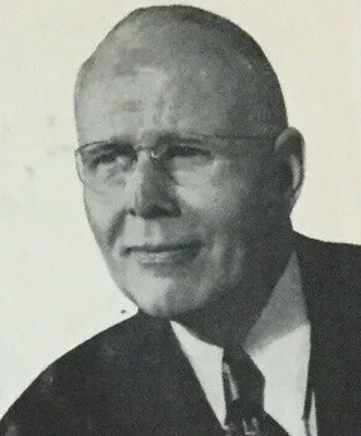
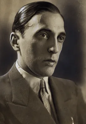
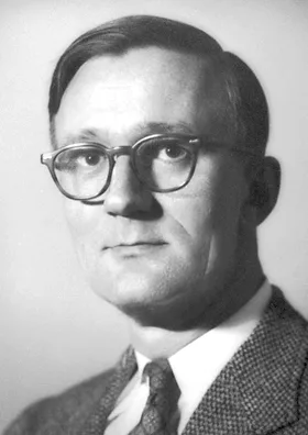
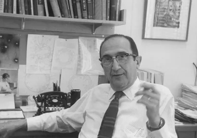
1932 ALVAREZ, Walter Clement (1884 – 1978); GIANTURCO, Cesare (1905 – 1995)
Roentgen ray motion pictures of the stomach.
Camera used for direct Roentgen-cinematography.
GMN 2697 · https://historyofmedicine.com/id/4308
1932 KNOLL, Max (1879 – 1969); RUSKA, Ernst August Friedrich (1906 – 1988)
Beitrag zur geometrischen Elektronenoptik.
Electron microscope. See also their later paper in Z. Physik. 1932, 78, 318.
GMN 269.3 · https://historyofmedicine.com/id/8749
1933 DELEE, Joseph Bolivar (1869 – 1942)
Sound motion pictures in obstetrics.
DeLee was one of the pioneers of filmmaking for the purpose of medical teaching. In this paper he described the necessary components of a medical film, including scripts, props, lighting, sound and expert staff member…
GMN 12485 · https://historyofmedicine.com/id/14718
1933 VAN DE VELDE, Theodoor Hendrik (1873 – 1937)
Sex efficiency through exercises. Special physical culture for women, with 480 cinematographic and 54 full-page illustrations.
Includes a series of 12 "cinematographic" films on 480 flicker cards. In all the photographs frontal nudity of the female model is covered by clothing, though her buttocks are exposed in some views. Having previously …
GMN 13200 · https://historyofmedicine.com/id/15461
1934 SNOW, William (1898 – ); POWELL, Clilian Bethany (1894 – 1977)
Roentgen visualization of the placenta.
Direct radiography of the placenta. "Clilan (C.B.) Powell, longtime owner of the Amsterdam News, was born in 1894 to former Virginia slaves. Very little is known about his childhood. He received his medical degree in …
GMN 6229 · https://historyofmedicine.com/id/7856
1934 BERNAL, John Desmond (1901 – 1971); HODGKIN, Dorothy Crowfoot (1910 – 1994)
X-Ray photographs of crystalline pepsin.
Bernal and Hodgkin took the first X-ray photograph of a protein structure—crystalline pepsin. They showed that crystals of pepsin give an X-ray diffaction pattern, beginning protein crystallography. This may also be t…
GMN 6923 · https://historyofmedicine.com/id/9088
1934 EGAS MONIZ, Antonio Caetano de Abreu Freire (1874 – 1955)
L'angiographie cérébrale, ses applications et résultats en anatomic, physiologie et clinique.
GMN 7766 · https://historyofmedicine.com/id/9938
1934 GESELL, Arnold Lucius (1880 – 1961)
An atlas of infant behavior: A systematic delineation of the forms and early growth of human behavior patterns... illustrated with 3,200 action photographs. Vol. l: Normative series, in collaboration with Helen Thompson and Catherine S. Amatruda. Selected bibliographies (p. 45). Vol. 2: Naturalistic series, in collaboration with Alice V. Keliher, Frances L. Ilg, and Jessie J. Carlson. (2 vols.)
Gesell, who originated the Child Study Center at Yale University, was the founder of the study of child development in the United States. He is best known for his groundbreaking studies of normal child development: be…
GMN 10609 · https://historyofmedicine.com/id/12802
1936 ABREU, Manuel Dias de (1894 – 1962)
Röntgen-photographia. Processo e apparelho de röntgen-photographia. Tuberculose pulmonar. Cadastra social. Radiographia e radioscopia. Röntgen-photographia collectiva.
Introduction of chest photofluorography or abreugraphy, also called mass miniature radiography.
GMN 3209.1 · https://historyofmedicine.com/id/4336
1937 ROESLER, Hugo (1899 – 1961)
Clinical roentgenology of the cardiovascular system.
GMN 2866 · https://historyofmedicine.com/id/4163
1938 RABI, Isidore Isaac (1898 – 1988); KUSCH, Polykarp (1911 – 1993); ZACHARAIS, Jerrold R. (1905 – 1986); ET AL
A new method of measuring nuclear magnetic moment.
Order of authorship in the original publication: Rabi, Zacharais,...Kusch. Followed by: (2) The magnetic moments of 3Li6, 3Li7, and 9F19. Physical Review 53, 1938. (3) The molecular beam resonance method for measuring…
GMN 14335 · https://historyofmedicine.com/id/16662
1942 LURIA, Salvador Edward (1912 – 1991); ANDERSON, Thomas Foxen (1911 – 1991)
The identification and characterization of bacteriophages with the electron microscope.
First identification, observation under the electron microscope, and reproduction of photomicrographs of bacteriophages taken through the electron microscope. Digital facsimile, but with poor reproductions of the two …
GMN 14004 · https://historyofmedicine.com/id/16309
1943 LURIA, Salvador Edward (1912 – 1991); DELBRÜCK, Max (1906 – 1981); ANDERSON, Thomas Foxen (1911 – 1991)
Electron microscope studies of bacterial viruses.
This work demonstrated the absorption of the phages on the host cell, and the lysis of the host cell and the liberation of a hundred or so daughter paritcles from each cell. It also showed that the particles multiply …
GMN 14005 · https://historyofmedicine.com/id/16310
1948 BRODÉN, Bror Leonhardt Johan (1910 – ); ET AL
Thoracic aortography. Preliminary report.
With H. E. Hanson and J. Karnell.
GMN 2879 · https://historyofmedicine.com/id/4220
1950 CASSEN, Benedict (1902 – 1972); ET AL
A sensitive directional gamma-ray detector.
Directional scintillation detector probe. Cassen assembled the first automated scanning system, comprised of a motor driven scintillation detector coupled to a relay printer. The scanner was initially used to image th…
GMN 2660.2 · https://historyofmedicine.com/id/3372
1951 ABEATICI, Sergio (1922 – 2000); CAMPI, L
La visualizzazione radiologica della porta pervia splenica.
Introduction of portal venography for investigation of portal hypertension.
GMN 2725.1 · https://historyofmedicine.com/id/4463
1951 COSSLETT, Vernon Ellis (1908 – 1990); NIXON, W. C.
X-ray shadow microscope.
GMN 269.7 · https://historyofmedicine.com/id/8752
1951 EHRENBERG, Werner (1901 – 1975); SPEAR, Walter Eric (1921 – 2008)
An electrostatic focusing system and its application to a fine focus x-ray tube.
X-ray microscopy.
GMN 269.8 · https://historyofmedicine.com/id/8753
1952 HOWRY, Douglas Hamilton (1920 – ); BLISS, Wiliam Roderic
Ultrasonic visualization of soft-tissue structures of the body.
First tomogram of soft tissue.
GMN 2680.1 · https://historyofmedicine.com/id/3567
1953 SELDINGER, Sven Ivar (1921 – 1998)
Catheter replacement of the needle in percutaneous angiography. A new technique.
Percutaneous arterial catheterization.
GMN 2924.2 · https://historyofmedicine.com/id/4927
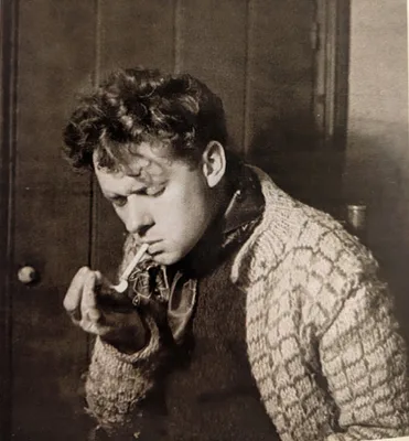
1953 THOMAS, Dylan (1914 – 1953)
The doctor and the devils.
The great lyric poet’s screenplay based on the notorious career of Robert Knox, the anatomist who purchased bodies for dissection from the resurrectionists/murderers, Burke and Hare. This was the first screenplay that…
GMN 6623.01 · https://historyofmedicine.com/id/7687
1954 EDLER, Inge Gudmar (1911 – 2001); HERTZ, Carl Hellmuth (1920 – 1990)
The use of ultrasonic reflectoscope for the continuous recording the movements of heart walls.
Echocardiography, from which the field of medical ultrasonics developed.
GMN 2883.01 · https://historyofmedicine.com/id/4242
1957 WILD, John Julian Cuttance (1914 – 2009); REID, John M. (1926 – )
Progress in the techniques of soft tissue examination by 15 MC pulsed ultrasound. In: Ultrasound in biology and medicine, ed. E. Kelly, pp. 30-48.
GMN 2681 · https://historyofmedicine.com/id/3578
1957 SATOMURA, Shigeo (1919 – 1960)
Ultrasonic Doppler method for the inspection of cardiac functions.
Demonstration of the Doppler shift in the frequency of ultrasound backscattered by moving cardiac structures.
GMN 2883.21 · https://historyofmedicine.com/id/4254
1958 DONALD, Ian (1910 – 1987)
Investigation of abdominal masses by pulsed ultrasound.
Donald, J. MacVicar and T. G. Brown used an ultrasound scanner to investigate the pregnant abdomen (see also No. 6235.1).
GMN 2682 · https://historyofmedicine.com/id/3582
1961 OLDENDORF, William Henry (1925 – 1992)
Isolated flying spot detection of radiodensity discontinuities displaying the internal structural pattern of a complex object.
Oldendorf described his experimental system for reconstructing the appearance of soft tissues by measuring radiodensity discontinuities (differences in tissue attenuation) but it was not fully recognized that very hig…
GMN 2700.3 · https://historyofmedicine.com/id/4337
1961 DONALD, Ian (1910 – 1987); BROWN, T. G.
Demonstration of tissue interfaces within the body by ultrasonic echo sounding.
Biparietal fetal cephalometry by ultrasound.
GMN 6235.1 · https://historyofmedicine.com/id/7872
1962 SONES, Frank Mason (1918 – 1985); SHIREY, Earl K. (1924 – )
Cine-coronary arteriography.
Coronary arteriography. Sones first performed this technique in 1958, and first published a more detailed account of it in a 1960 book chapter: "Cinecardioangiography," pp. 130-144 in B. L. Gordon (ed.) Clinical cardi…
GMN 2924.3 · https://historyofmedicine.com/id/4929
1963 CORMACK, Allan McLeod (1924 – 1998)
Representation of a Function by its Line Integrals, with Some Radiological Applications.
Cormack showed that changes in tissue density could be computed from x-ray data. Because of limitations in computing power no machine was constructed during the 1960s. Cormack's papers generated little interest until …
GMN 8149 · https://historyofmedicine.com/id/10325
1965 THOMPSON, Horace E. (1921 – 2008); HOLMES, Joseph H. (1902 – 1982); GOTTESFELD, Kenneth R.; TAYLOR, E. S.
Fetal development as determined by ultrasonic pulse echo techniques.
Order of authorship in the original publication: Thompson, Holmes, Gottesfeld, Taylor. This was the first paper on the use of ultrasound in obstetrics and gynecology published in the United States.
GMN 11463 · https://historyofmedicine.com/id/13662
1965 MIYAMOTO, Kaneatsu; MATSUMOTO, Seiichi
The nature of the Negri Body.
The first visual proof, by publication of electron micrographs at 25,000 magnification, that Negri bodies contain enormous numbers of rabies virus particles. Digital facsimile from rupress.org at this link. (Thanks to…
GMN 14035 · https://historyofmedicine.com/id/16343
1967 JUDKINS, Melvin Paul (1922 – 1985)
Selective coronary arteriography. Part I: A percutaneous transfemoral technic.
Selective percutaneous transfemoral arteriography.
GMN 2924.5 · https://historyofmedicine.com/id/4933
1968 KLUG, Sir Aaron (1926 – 2018); DEROSIER, David J.
Reconstruction of three-dimensional structures from electron micrographs.
Klug and deRosier invented methods for two-dimensional and three-dimensional digital image processing of electron microscope images. The latter method provided the theory behind the development of computed tomography …
GMN 13935 · https://historyofmedicine.com/id/16231
1973 HOUNSFIELD, Sir Godfrey Newbold (1919 – 2004)
Computerized transverse axial scanning (tomography).
Hounsfield invented computer-assisted tomography (CAT), or computed tomography (CT). In 1979 Hounsfield shared the Nobel Prize in Physiology or Medicine with Allen M. Cormack "for the development of computer assisted …
GMN 2700.4 · https://historyofmedicine.com/id/4340
1973 LAUTERBUR, Paul Christian (1929 – 2007)
Image formation by induced local interactions: Examples employing nuclear magnetic resonance.
Lauterbur proposed a workable method for using nuclear magnetic resonance to produce images of tissues. In 2003 Lauterbur shared the Nobel Prize in Physiology or Medicine with Sir Peter Mansfield “for their discoverie…
GMN 2700.5 · https://historyofmedicine.com/id/4343
1974 LEDLEY, Robert Steven (1926 – 2012); ET AL
Computerized transaxial x-ray tomography of the human body.
Ledley and team developed the developed the ACTA 0100 CT Scanner (Automatic Computerized Traverse Axial)— the first whole-body computed tomography scanner. With G. Di Chiro, A. J. Luessenhop, and H.L. Twigg. For furth…
GMN 6909 · https://historyofmedicine.com/id/9073
1976 GILMAN, Sander L. (1941 – ); DIAMOND, Hugh Welch (1809 – 1886)
The face of madness. Hugh W. Diamond and the origin of psychiatric photography. Edited by Sander L. Gilman.
Papers by Diamond, including their illustrations, edited with an extensive annotated introduction.
GMN 7072 · https://historyofmedicine.com/id/9239
1977 MANSFIELD, Sir Peter (1933 – 2017); MAUDSLEY, Andrew A.
Medical imaging by NMR.
Mansfield developed a mathematical technique that would allow NMR scans to take seconds rather than hours and produce clearer images than the technique Paul Lauterbur developed in 1973. Mansfield showed how gradients …
GMN 8150 · https://historyofmedicine.com/id/10326
1978 HATLE, Liv Kristin (1936 – ); ANGELSEN, Bjorn A. J. (1948 – ); BRUBAKK, Alf O.; TROMSDAL, Arve
Noninvasive assessment of pressure drop in mitral stenosis by Doppler ultrasound.
Order of authorship in the original paper: Hatle, Brubakk, Tromsdal, Angelsen. "Hatle (born 1936) was the pioneer of continuous wave Doppler echocardiography. "Today, Doppler echocardiography is central to our ability…
GMN 12243 · https://historyofmedicine.com/id/14457
1986 NIELSEN, Arnold B. W.; RØNNE, Paul
Development of the ion X-ray tube: With a description of the Collection in the Medical Historical Museum, University of Copenhagen.
GMN 13185 · https://historyofmedicine.com/id/15446
1990 DIAMOND, Hugh Welch (1809 – 1886)
Portraits of the insane. The case of Dr. Diamond, by Adrienne Burrows and Iwan Schumacher.
Reproduces many of Diamond's photographs of psychiatric patients. Diamond was fascinated by the possible use of photography in the treatment of mental disorders; some of his many photographs depicting the expressions …
GMN 7073 · https://historyofmedicine.com/id/9240
1990 WEBB, Steve
From the watching of shadows. The origins of radiological tomography.
GMN 7180 · https://historyofmedicine.com/id/9347
1991 ELLIS, Harold (1926 – ); LOGAN, Bari M.; DIXON, Adrian Kendall (1948 – )
Human cross-sectional anatomy: Atlas of body sections and CT images.
GMN 7718 · https://historyofmedicine.com/id/9890
1994 ROSENBUSCH, Gerd; OUDKERK, Matthijs; AMMANN, Ernst
Radiologie in der medizinischen Diagnostik. Evolution der Röntgenstrahlenanwendung 1895-1995.
Includes a section on the development of tomography and computed tomography (CT). Translated into English by Peter F. Winter as Radiology in medical diagnostics: Evolution of X-ray applications 1895-1995 (Oxford: Blac…
GMN 7583 · https://historyofmedicine.com/id/9755
1996 MATTSON, James; SIMON, Merrill
The pioneers of NMR and magnetic resonance in medicine: The story of MRI.
GMN 7185 · https://historyofmedicine.com/id/9352
1996 BENGSTON, Bradley P.; KUZ, Julian E.
Photographic atlas of Civil War injuries. Photographs of surgical cases and specimens. Otis Historical Archives.
GMN 9426 · https://historyofmedicine.com/id/11609
1996 BENGSTON, Bradley P.; KUZ, Julian E.
Orthopaedic injuries of the Civil War: An atlas of orthopaedic injuries and treatments during the Civil War.
GMN 9427 · https://historyofmedicine.com/id/11610
1997 KEVLES, Bettyann Holtzmann (1938 – )
Naked to the bone. Medical imaging in the twentieth century.
GMN 7181 · https://historyofmedicine.com/id/9348
1999 PERNICK, Martin S. (1948 – )
The black stork: Eugenics and the death of "defective" babies in American medicine and motion pictures since 1915.
GMN 10053 · https://historyofmedicine.com/id/12242
2002 MCGRATH, Roberta
Seeing her sex: Medical archives and the female body.
"Through a detailed analysis of exterior and interior images of the female body, this book examines the relationship between human reproduction and cultural representation from 1750-1910. With examples drawn from medi…
GMN 10175 · https://historyofmedicine.com/id/12364