1895 RÖNTGEN, Wilhelm Conrad (1845 – 1923)
Ueber eine neue Art von Strahlen. (Vorläufige Mittheilung.); Eine neue Art von Strahlen. II. Mittheilung.
The discovery of x rays, which Kölliker later renamed “Roentgen rays”; the foundation stone of the science of roentgenology. English translation in Nature, (February,) 1896, 53, 274 and 377. See also Röntgen’s third p…
GMN 2683 · https://historyofmedicine.com/id/4359
1896 WILLIAMS, Francis Henry (1852 – 1936)
A method for more fully determining the outline of the heart by means of the fluoroscope together with other uses of this instrument in medicine.
Estimation of heart size by fluoroscope, first application of x rays to cardiology.
GMN 2804.1 · https://historyofmedicine.com/id/3439
1896 BECHER, Wolf (1862 – 1906)
Zur Anwendung des Röntgenschen Verfahrens in der Medicin.
Becher introduced a solution of lead into the stomach of a guinea-pig, making it opaque to x rays; he thus showed the possibility of radiological diagnosis of gastric disease.
GMN 3516 · https://historyofmedicine.com/id/3992
1896 JONES, Sir Robert (1858 – 1933); LODGE, Sir Oliver Joseph (1851 – 1940)
The discovery of a bullet lost in the wrist by means of the Roentgen rays.
This was probably the first published report of the clinical use of x rays (February 22).
GMN 2684 · https://historyofmedicine.com/id/4167
1896 THOMSON, Elihu (1853 – 1937)
Stereoscopic Roentgen pictures.
Invention of the Roentgen stereoscope.
GMN 2686 · https://historyofmedicine.com/id/4204
1896 WILLIAMS, Francis Henry (1852 – 1936)
A method for more fully determining the outline of the heart by means of the fluorescope together with other uses of this instrument in medicine.
Estimation of heart size by fluoroscope, first application of x rays to cardiology.
GMN 2686.1 · https://historyofmedicine.com/id/4210
1896 CHAPPUIS, J; CHAUVEL, H
Calculs du rein. Calculs de la vésicule biliaire.
Chappuis and Chauvel were the first to study biliary concretions by means of Roentgen rays.
GMN 3636 · https://historyofmedicine.com/id/4634
1896 MACINTYRE, John (1857 – 1928)
Roentgen rays. Photography of renal calculus.
First radiogram of renal calculus.
GMN 4293 · https://historyofmedicine.com/id/5383
1896 HARRISON, Frank (1859 – 1912)
The “x” rays in the practice of dental surgery.
Harrison was the first to describe a method of making dental radiographs.
GMN 3688 · https://historyofmedicine.com/id/5699
1896 KÖNIG, Carl Georg Walter (1859 – 1936)
14 Photographien mit Röntgen-strahlen aufgenommen im physikaloschen Verein zu Frankfurt A. M.
This collection of x-ray photographs published within a few months of Röntgen's discovery includes applications in archaeology and anthropology (x-rays of mummies) and forensic medicine (for the investigation of gunsh…
GMN 10608 · https://historyofmedicine.com/id/12801
1897 MACINTYRE, John (1857 – 1928)
X-ray records for the cinematograph.
Macintyre was the first to demonstrate x-ray cinematography.
GMN 2687 · https://historyofmedicine.com/id/4216
1898 BUXBAUM, A
Ueber die Photographie von Gallensteinen in vivo.
First x-ray demonstration of gall-stones.
GMN 3638 · https://historyofmedicine.com/id/4636
1899 TUFFIER, Théodore (1857 – 1929)
Sonde urétérale opaque. In Traité de chirurgie, 2e ed. vol. 7, p. 414.
Tuffier made the first pioneer attempt to visualize the urinary tract by the combination of an opaque ureteral styletted catheter and radiography.
GMN 4191.1 · https://historyofmedicine.com/id/6119
1901 ILLYÉS, Géza (1870 – 1951)
Uretercatheterezés és radiographia.
Géza von Illyés showed that ureteral calculi could be accurately demonstrated by x rays with the help of an indwelling opaque catheter. German translation in Dtsch. Z. Chir., 1901, 62, 132-40.
GMN 4294 · https://historyofmedicine.com/id/5388
1902 MORITZ, Friedrich (1861 – 1938)
Ueber orthodiagraphische Untersuchungen am Herzen.
Orthodiagraphy of the heart.
GMN 2813 · https://historyofmedicine.com/id/3479
1903 –1904 ALBERS-SCHÖNBERG, Heinrich Ernst (1865 – 1921)
Projektions-Röntgenbilder einer seltenen Knochenerkrankung.
First description of osteosclerosis fragilis, marble bones (“Albers-Schönberg disease”).
GMN 4372 · https://historyofmedicine.com/id/5858
1905 KÖHLER, Alban (1874 – 1947)
Technik der Herstellung fast orthodiagraphischer Herzphotogramme vermittelst Röntgeninstrumentarien mit kleiner Elektrizitätsquelle.
Introduction of teleradiography of the heart.
GMN 2817 · https://historyofmedicine.com/id/3496
1905 VOELCKER, Friedrich (1872 – 1955); LICHTENBERG, Alexander von (1880 – 1949)
Die Gestalt der menschlichen Harnblase im Röntgenbilde.
First cystograms.
GMN 4192 · https://historyofmedicine.com/id/6129
1908 KUTTNER, Arthur (1862 – 1939)
Die entzündlichen Nebenhöhlenerkrankungen der Nase im Röntgenbild.
The first important work on the radiology of the accessory nasal sinuses.
GMN 3316 · https://historyofmedicine.com/id/4532
1909 GROEDEL, Franz Maximilian (1881 – 1951)
Die Technik der Roentgenkinematographie.
Groedel invented the first machine for taking serial x-rays.
GMN 2690 · https://historyofmedicine.com/id/4269
1911 EDLING, Lars (1878 – 1962)
Ueber die Anwendung des Roentgenverfahrens bei der Diagnose der Schwangerschaft.
First use of x rays for the diagnosis of pregnancy.
GMN 6216 · https://historyofmedicine.com/id/7632
1913 HUISMANS, L
Der Ersatz des Orthiodiagraphen durch der Teleröntgen.
Instantaneous radiography of the heart.
GMN 2842 · https://historyofmedicine.com/id/3595
1916 CRANE, Augustus Warren (1868 – 1937)
Roentgenology of the heart.
Introduction of kymography in clinical cardiology.
GMN 2845 · https://historyofmedicine.com/id/3609
1917 ROWNTREE, Leonard George (1883 – 1959); WATERS, Charles Alexander (1888 – 1961); BAYNE-JONES, Brig. Gen. Stanhope (1866 – 1970)
Roentgenography of the lung: Roentgenographic studies in living animals after intratracheal injection of iodoform emulsion.
Experimental introduction of iodoform (lipiodol) into the bronchial tree in dogs, obtaining satisfactory bronchograms.
GMN 3194.1 · https://historyofmedicine.com/id/4252
1917 THOMA, Kurt Hermann (1883 – 1972)
Oral roentgenology.
GMN 3690 · https://historyofmedicine.com/id/5716
1918 DANDY, Walter Edward (1886 – 1946)
Ventriculography following the injection of air into the cerebral ventricles.
Dandy was responsible for the introduction of ventriculography.
GMN 4602 · https://historyofmedicine.com/id/8927
1919 DANDY, Walter Edward (1886 – 1946)
Röntgenography of the brain after the injection of air into the spinal canal.
Introduction of pneumoencephalography.
GMN 4603 · https://historyofmedicine.com/id/554
1920 YOUNG, Hugh Hampton (1870 – 1945); WATERS, Charles Alexander (1888 – 1961)
X-ray studies of the seminal vesicles and vasa deferentia after urethroscopic injection of the ejaculatory ducts with thorium – a new diagnostic method.
Vesiculography first demonstrated.
GMN 4198 · https://historyofmedicine.com/id/6159
1921 FORESTIER, Jacques (1890 – 1978); SICARD, Jean Athanase (1872 – 1929)
Méthode radiographique d’exploration de la cavité épidurale par la lipiodol.
Positive contrast myelography with iodized oil (lipiodol). This paper records the first use of lipiodol in radiology.
GMN 2693 · https://historyofmedicine.com/id/556
1921 WIDERØE, Sofus (1880 – 1937)
Über die diagnostische Bedeutung der intraspinalen Luftinjektionen bei Rückenmarksleiden, besonders bei Geschwülsten.
Myelography by air injection into spinal subarachnoid space.
GMN 4605.1 · https://historyofmedicine.com/id/558
1921 LYNAH, Henry Lowndes (1879 – 1922); STEWART, William Holmes (1869 – 1954)
Roentgenographic studies of bronchiectasis and lung abscess after direct injection of bismuth mixture through the bronchoscope.
Important studies on bronchiectasis were carried out by Lynah and Stewart.
GMN 3197 · https://historyofmedicine.com/id/4270
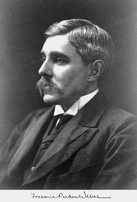
1922 WEBER, Frederick Parkes (1863 – 1962)
Right-sided hemi-hypertrophy resulting from right-sided congenital spastic hemiplegia, with a morbid condition of the left side of the brain, revealed by radiograms.
Sturge–Weber syndrome (see No. 4560.1).
GMN 4605.2 · https://historyofmedicine.com/id/564
1923 BERBERICH, Josef (1897 – ); HIRSCH, S
Die Röntgenographische Darstellung der Arterien und Venen am lebenden Menschen.
Arteriography. First angiogram of a living patient.
GMN 2916 · https://historyofmedicine.com/id/4895
1923 ROWNTREE, Leonard George (1883 – 1959); OSBORNE, Earl Dorland (1895 – 1960); ET AL
Roentgenography of the urinary tract during excretion of sodium iodide.
First use of sodium iodide in uretero-pyelography. With C. G. Sutherland and A. J. Scholl.
GMN 4199 · https://historyofmedicine.com/id/6166
1924 FORESTIER, Jacques (1890 – 1978); SICARD, Jean Athanase (1872 – 1929)
L’exploration radiologique des cavités broncho-pulmonaires par les injections intra-trachéales d’huile iodée.
Bronchography was advanced by the work of Sicard and Forestier on the intratracheal introduction of lipiodol.
GMN 3199 · https://historyofmedicine.com/id/4292
1924 GRAHAM, Evarts Ambrose (1883 – 1957); COLE, Warren Henry (1898 – 1990)
Roentgenologic examination of the gallbladder.
Introduction of cholecystography.
GMN 3652 · https://historyofmedicine.com/id/4751
1924 BROOKS, Barney (1884 – 1952)
Intra-arterial injection of sodium iodide: Preliminary report.
Clinical angiography.
GMN 2916.1 · https://historyofmedicine.com/id/4897
1924 BROOKS, Barney (1884 – 1952); JOSTES, F. A.
A clinical study of diseases of the circulation of the extremities; a description of a new method of examination.
Femoral arteriography.
GMN 2916.2 · https://historyofmedicine.com/id/4898
1925 RAPER, Howard Riley (1887 – 1978)
A new kind of x-ray examination for preventive dentistry.
Original description of technique of making “bite-wing” radiographs.
GMN 3692 · https://historyofmedicine.com/id/5731
1927 EGAS MONIZ, Antonio Caetano de Abreu Freire (1874 – 1955)
L’encéphalographie artérielle, son importance dans la localisation des tumeurs cérébrales.
Introduction of cerebral arteriography. See also Presse méd., 1928, 36, 689-93. English translation in J. Neurosurg., 1964, 21, 145-56.
GMN 4610 · https://historyofmedicine.com/id/578
1927 EGAS MONIZ, Antonio Caetano de Abreu Freire (1874 – 1955)
Injections intracarotidiennes et substances injectables opaques aux rayons X.
Carotid arteriography.
GMN 4610.1 · https://historyofmedicine.com/id/8929
1930 MAYER, Ernst Georg (1893 – )
Otologische Röntgendiagnostik.
Includes a brief history of the subject.
GMN 3407 · https://historyofmedicine.com/id/5208
1930 MENEES, Thomas Orville (1890 – 1937); ET AL
Amniography; preliminary report.
Introduction of amniography. With J. D. Miller and L. E. Holly.
GMN 6223 · https://historyofmedicine.com/id/7649
1931 PEARSE, Herman Elwyn (1899 – ); WARREN, Stafford Leak (1896 – 1981)
The roentgenographic visualization of the arteries of the extremities in peripheral vascular disease.
GMN 2919.1 · https://historyofmedicine.com/id/4906
1931 SANTOS, Reynaldo dos (1880 – 1970); CALDAS, Jose Guilherme Mendes Pereira
Les dérivés du thorium dans l’artériographie des membres.
Thorotrast first used in arteriography.
GMN 2920 · https://historyofmedicine.com/id/4909
1931 LYSHOLM, Erik (1891 – 1947)
Apparatus and technique for roentgen examination of the skull.
Lysholm–Schönander skull table, allowing precise radiography of the skull.
GMN 4611.1 · https://historyofmedicine.com/id/8930
1931 WALKHOFF, Friedrich Otto (1860 – 1934)
Das Problem der dentalen Fokalinfektion und ihre Bekämpfung durch die konservierende Zahnheilkunde.
With physicist Fritz Giesel, Walkhoff on December 28, 1895 Walkhoff did the first dental radiograph on his own teeth. Later, Walkhoff was "responsible for the shift to an everyday use of camphorated chlorophenol to st…
GMN 12985 · https://historyofmedicine.com/id/15233
1932 ALVAREZ, Walter Clement (1884 – 1978); GIANTURCO, Cesare (1905 – 1995)
Roentgen ray motion pictures of the stomach.
Camera used for direct Roentgen-cinematography.
GMN 2697 · https://historyofmedicine.com/id/4308
1932 KNOLL, Max (1879 – 1969); RUSKA, Ernst August Friedrich (1906 – 1988)
Beitrag zur geometrischen Elektronenoptik.
Electron microscope. See also their later paper in Z. Physik. 1932, 78, 318.
GMN 269.3 · https://historyofmedicine.com/id/8749
1934 SNOW, William (1898 – ); POWELL, Clilian Bethany (1894 – 1977)
Roentgen visualization of the placenta.
Direct radiography of the placenta. "Clilan (C.B.) Powell, longtime owner of the Amsterdam News, was born in 1894 to former Virginia slaves. Very little is known about his childhood. He received his medical degree in …
GMN 6229 · https://historyofmedicine.com/id/7856