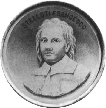
1630 STELLUTI, Francesco (1577 – 1653)
Persio tradotto.
The first book to contain illustrations of natural objects as seen through the microscope— specifically an engraving of the exterior surface of bees. The work includes the Latin text of the Satyrae VI of Aulus Persius…
GMN 259 · https://historyofmedicine.com/id/8731
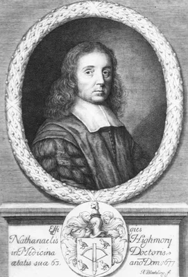
1651 HIGHMORE, Nathaniel (1613 – 1685)
The history of generation…
Highmore’s account of the development of the chick is the first embryological study based on microscopical examination, predating Malpighi (No. 468) by more than twenty years. This is also the first book in English to…
GMN 467.1 · https://historyofmedicine.com/id/1268
1653 BOREL, Pierre [BORELLUS.] (1620 – 1689)
Historiarum et observationum medico-physicarum centuria prima (-secunda).
The first work to apply microscopy to medicine. Borel described 200 observations and applications; he probably saw the blood corpuscles and Sarcoptes scabiei.
GMN 260 · https://historyofmedicine.com/id/8733
1655 BOREL, Pierre [BORELLUS.] (1620 – 1689)
De vero telescopii inventore.
Borel collected evidence to show that Zacharias (sometimes called Zacharias Janssen) invented the compound microscope about 1590. Zacharias was a spectacle-maker of Middelburg, Holland.
GMN 261 · https://historyofmedicine.com/id/8734
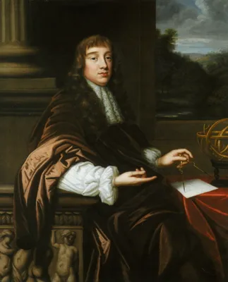
1665 HOOKE, Robert (1635 – 1703)
Micrographia, or some physiological descriptions of minute bodies made by magnifying glasses; with observations and inquiries thereupon.
Hooke, at one time research assistant to Robert Boyle, was one of the greatest inventive geniuses of all time. This was the first book devoted entirely to microscopical observations, and also the first book to pair it…
GMN 262 · https://historyofmedicine.com/id/8735
1678 HOOKE, Robert (1635 – 1703)
Microscopium, or some new discoveries made with and concerning microscopes. Lectures and Collections made by Robert Hooke, Secretary of the Royal Society, pages 81-112.
On his title page Hooke listed the contents of this section as follows: "Mr. Leeuwenhoeck's two letters concerning some late microscopical discoveries. "The author's discourse and description of microscopes, improved …
GMN 13233 · https://historyofmedicine.com/id/15497
1685 –1686 ZAHN, Johann (1641 – 1707)
Oculus artificialis teledioptricus sive telescopium. 3 pts.
Includes the first complete history of early microscopes.
GMN 263 · https://historyofmedicine.com/id/8736
1686 –1718 LEEUWENHOEK, Antonie van (1632 – 1723)
Ontledingen en ontdekkingen.... 6 vols.
Leeuwenhoek, one of the first and also one of the greatest of the microbiologists, communicated many of his discoveries to the Royal Society in London. This set is a collection in Dutch of many contributions that van …
GMN 265 · https://historyofmedicine.com/id/1222
1691 BONANNI, Filippo [BUONANNI] (1638 – 1723)
Observationes circa viventia, quae in rebus non viventibus reperiuntur. Cum micrographia curiosa siue Rerum minutissimarum obseruationibus, quæ ope microscopij recognitæ ad viuum exprimuntur. His accesserunt aliquot animalium testaceorum icones non antea in lucem editae. Omnia curiosorum naturæ exploratorum vtilitati & iucunditati expressa & oblata.
Illustrates several early microscopes, including the famous microscopes of the Bolognese Joseph Campani. Contradicting Redi, Bonanni tried to show that spontaneous generation was possible in animals "without blood and…
GMN 264 · https://historyofmedicine.com/id/8737
1762 LYONET, Pierre (1707 – 1789)
Traité anatomique de la chénille, qui ronge le bois de saule. Augmenté d'une explication abrégée des planches, et d'une description de l'instrument et des outils dont l'auteur s'est servi pour anatomiser à la loupe et au microscope, & pour déterminer la forcer de ses verres, suivant les règles de l'optique, & méchaniquement.
Lyonet’s monograph on the goat moth caterpillar remains a famous example of anatomical examination. It is also a thorough treatise on the microscope and lenses that Lyonet used. Digital facsimile from Biodiversity Her…
GMN 305 · https://historyofmedicine.com/id/239
1807 DEIJL, Hermanus van (1738 – 1809)
Kort bericht der trapsgewijze verbeteringen aan achromatische verrekijkers.
Van Deijl introduced an achromatic objective.
GMN 265.1 · https://historyofmedicine.com/id/8739
1820 AMICI, Giovanni Battista (1784 – 1863)
De' microscopj catadiottrici memoria.
Amici constructed the first microscope with achromatic lenses and suggested water-immersion for improved achromatic lenses of the compound microscope. The 1820 publication of his paper indicates that it was "Approvata…
GMN 266 · https://historyofmedicine.com/id/8740
1827 GORING, C. R. (1792 – 1840)
On Mr. Tulley’s thick aplanatic object-glasses, for diverging rays; with an account of a few microscopic test objects.
Goring was an Edinburgh medical practitioner. He commissioned Tulley and others to make various modifications to the microscope. The above paper reports the first effective achromatic object-glass.
GMN 266.1 · https://historyofmedicine.com/id/8741
1827 HODGKIN, Thomas (1798 – 1866); LISTER, Joseph Jackson (1786 – 1869)
Notice of some microscopic observations of the blood and animal tissues.
Hodgkin and Lister determined that blood cells are bioconcave discs and accurately measured their diameter as "1/5000 of an inch." Digital facsimile from Google Books at this link.
GMN 13262 · https://historyofmedicine.com/id/15528
1830 LISTER, Joseph Jackson (1786 – 1869)
On some properties in achromatic object-glasses applicable to the improvement of the microscope.
The principle of the modern microscope was worked out by Joseph Jackson Lister, father of Lord Joseph Lister. His important improvements in achromatic lenses make him one of the most prominent figures in the history o…
GMN 267 · https://historyofmedicine.com/id/8742
1838 –1857 MANDL, Louis (1812 – 1881)
Anatomie microscopique. Tome premier: Histologie. Atlas de cinquante-deux planches. Tome second: Histogénèse. Atlas de quarante planches. 2 vols.
This work was issued in parts over nearly 20 years. Parts concerned specific subjects and were sold separately. Digital facsimile from Google Books at this link, and at this link.
GMN 11117 · https://historyofmedicine.com/id/13313
1844 –1845 DONNÉ, Alfred François (1801 – 1878); FOUCAULT, Jean Bernard Léon (1819 – 1868)
Cours de microscopie. 1 vol. and atlas.
Donne's and Foucault's work was the first biomedical textbook to be illustrated with images made from photomicrographs, in this case daguerreotypes of blood cells. Among its noteworthy images are the first microphotog…
GMN 267.1 · https://historyofmedicine.com/id/8743
1848 –1854 HARTING, Pieter (1812 – 1885)
Het mikroskoop, deszelfs gebruik, Geschiedenis en tegewoordige Toestand. Eeen handbooek voor natuur - en geneeskundigen. 4 vols.
Exhaustive history of the microscope. The work was translated into German, appearing (second edition) in 1866; this last was reprinted Amsterdam, 3 vols., 1970. Digital facsimile of the first edition in Dutch from wel…
GMN 270 · https://historyofmedicine.com/id/8754
1870 HIS, Wilhelm Sr. (1831 – 1904)
Beschreibung eines Mikrotoms.
His was, more than any other person, responsible for the introduction of the microtome, although Ranvier and other French people had earlier employed microtomes of simpler types.
GMN 268 · https://historyofmedicine.com/id/8744
1878 STEPHENSON, John Ware
On a large-angled immersion objective, without adjustment collar; with some observations on “numerical aperture”.
Stephenson suggested the oil immersion lens system to Abbe, who developed it.
GMN 268.1 · https://historyofmedicine.com/id/8745
1886 ABBE, Ernst Karl (1840 – 1905)
Ueber neue Mikroskope.
Fundamental improvements in the microscope were made by Abbe, who was a mathematician. In 1878 he introduced the oil immersion lens; in 1886 he made an apochromatic objective corrected for three colors in which the se…
GMN 269 · https://historyofmedicine.com/id/8746
1902 BORST, Maximilian (1869 – 1946)
Die Lehre von den Geschwülsten. 2 vols.
“With this book the microscopical epoch in the evolution of the knowledge of cancer may be said to have been brought to a close” (Haagensen).
GMN 2625 · https://historyofmedicine.com/id/3179
1904 KÖHLER, August Karl Johann Valentin (1866 – 1948)
Mikrophotographische Untersuchungen mit ultraviolettem Licht.
The ultraviolet light microscope was conceived and designed by Köhler.
GMN 269.1 · https://historyofmedicine.com/id/8747
1911 HEIMSTÄDT, Oskar (1879 – 1944)
Das Fluoreszenzmikroskop.
Fluorescence microscopy
GMN 269.2 · https://historyofmedicine.com/id/8748
1918 –1973 PURKYNĔ, Jan Evangelista [PURKINJE] (1787 – 1869)
Sebrané spisy. Opera omnia. Tom. 1-12.
Purkynĕ was Professor of Physiology at Breslau and Prague. Eminent as physiologist and microscopist, he was first to use the microtome. See Kruta, V. J.E. Purkynĕ, Physiologist. A short account of his contributions…wi…
GMN 82 · https://historyofmedicine.com/id/1378
1921 VOGT, Alfred (1879 – 1943)
Atlas der Spaltlampenmikroskopie des lebenden Auges.
An important work on the biomicroscopy of the eye. Second ed. greatly revised and enlarged, vol. 1-2, Springer, 1930-31; vol. 3, Stuttgart, F. Enke, 1942; vol. 3 (English translation) Zurich, 1947. Second ed. reprinte…
GMN 1527 · https://historyofmedicine.com/id/2853
1932 KNOLL, Max (1879 – 1969); RUSKA, Ernst August Friedrich (1906 – 1988)
Beitrag zur geometrischen Elektronenoptik.
Electron microscope. See also their later paper in Z. Physik. 1932, 78, 318.
GMN 269.3 · https://historyofmedicine.com/id/8749
1932 CLAY, Reginald Stanley (1868 – 1964); COURT, Thomas H.
The history of the microscope.
A classic history of microscopes up to 1800.
GMN 271 · https://historyofmedicine.com/id/8790
1934 ABDERHALDEN, Emil (1877 – 1950); HAITINGER, Max (1868 – 1946)
Die Methoden der Fluoreszenzmikroskopie. In: Abderhalden, Handbuch der biologischen Arbeitsmethoden, Abt. II, Teil 3, pp. 3307-37.
Modern methods of fluorescence microscopy were developed by Haitinger.
GMN 269.4 · https://historyofmedicine.com/id/8825
1935 ZERNIKE, Frits (1888 – 1966)
Das Phasenkontrastverfahren b.d. mikroskopischen Beobachtung.
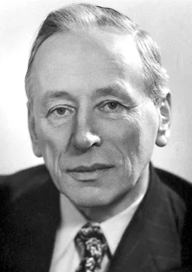
Zernicke invented phase contrast microscopy. In 1953 Zernike was awarded the Nobel Prize in Physics "for his demonstration of the phase contrast method, especially for his invention of the phase contrast microscope."
GMN 269.5 · https://historyofmedicine.com/id/8750
1936 BARNARD, Joseph Edwin (1870 – 1949); WELCH, Frank V.
Microscopy with ultra-violet light. A simplification of method.
GMN 269.6 · https://historyofmedicine.com/id/8751
1939 –2015 LEEUWENHOEK, Antonie van (1632 – 1723)
The collected letters of Antoni Van Leeuwenhoek / Alle de Brieven van Antoni Van Leeuwenhoek. Edited, illustrated & annotated by a committee of Dutch scientists. 17 vols. through 2015.
GMN 12964 · https://historyofmedicine.com/id/15211
1950 DYSON, J.
An inferometer microscope.
Dyson designed one of the first usable interference microscopes This optical system achieved interference imaging without requiring polarizing elements in the beam path.
GMN 7568 · https://historyofmedicine.com/id/9740
1951 COSSLETT, Vernon Ellis (1908 – 1990); NIXON, W. C.
X-ray shadow microscope.
GMN 269.7 · https://historyofmedicine.com/id/8752
1951 EHRENBERG, Werner (1901 – 1975); SPEAR, Walter Eric (1921 – 2008)
An electrostatic focusing system and its application to a fine focus x-ray tube.
X-ray microscopy.
GMN 269.8 · https://historyofmedicine.com/id/8753
1963 –1966 FREUND, Hugo; BERG, Alexander
Geschichte der Mikroskopie. 3 vols.
A biographical history.
GMN 273 · https://historyofmedicine.com/id/8792
1967 BRADBURY, Savile (1931 – 2001)
The evolution of the microscope.
GMN 273.1 · https://historyofmedicine.com/id/8794
1968 MARTON, Ladislaus Laslo (1901 – 1979)
Early history of the electron microscope.
GMN 273.2 · https://historyofmedicine.com/id/8795
1971 HOGAN, Michael J. (1907 – 1976); ALVARADO, Jorge A.; WEDDELL, Joan Esperson
Histology of the human eye.
Hogan and Alvarado's work was the first book on histology of the eye to include electron microscopy. It also reproduced spectacular three-dimensional representations of ocular ultrastructures by Joan Esperson Weddell.
GMN 7405 · https://historyofmedicine.com/id/9577
1975 HENDERSON, Richard (1945 – ); UNWIN, Peter Nigel Tripp
Three-dimensional model of purple membrane obtained by electron microscopy.
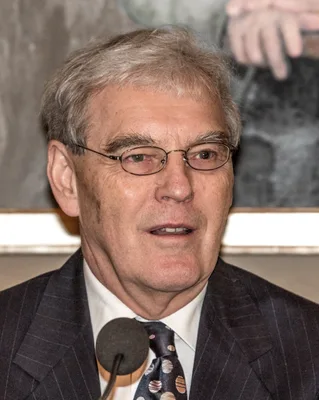
The invention of Cryogenic electron microscopy (cryo-EM). The novel technique was achieved by "by applying the method to tilted specimens, and using the principles put forward by De Rosier and Klug (GM - 13935), for t…
GMN 14182 · https://historyofmedicine.com/id/16497
1978 BRACEGIRDLE, Brian (1933 – 2015)
A history of microtechnique: The evolution of the microtome and the development of tissue preparation.
GMN 567.2 · https://historyofmedicine.com/id/320
1981 FRANK, Joachim (1940 – ); VERSCHOOR, Adriana; BOUBLIK, Miloslav (1929 – 1994)
Computer averaging of electron micrographs of the 405 ribosomal subunit.
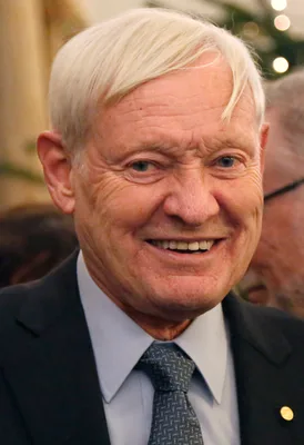
Frank and colleagues developed a method that allows sorting of particle images into classes based on their orientation, as well as their structural features. Specifically Frank developed mathematical tools used for im…
GMN 14183 · https://historyofmedicine.com/id/16498
1983 CONN, Harold Joel (1886 – 1975); KASTEN, Frederick H. (1927 – 2014); LUSK, George C.
History of staining by Harold J. Cohn. 3rd edition by George C. Lusk and Frederick H.Kasten.
GMN 272 · https://historyofmedicine.com/id/8803
1984 DUBOCHET, Jacques (1942 – ); ADRIAN, Marc; LEPAULT, Jean; ET AL
Cryo-electron microscopy of viruses.
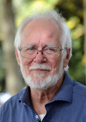
Dubochet and colleagues introduced "Dubochet's vitrification method" to vitrify water by cooling it so rapidly that it solidified to form a glass instead of crystals. Using this method, the authors published the first…
GMN 14184 · https://historyofmedicine.com/id/16499
1989 TURNER, Gerard l'Estrange
The great age of the microscope: The collection of the Royal Microscopical Society through 150 years.
GMN 13311 · https://historyofmedicine.com/id/15577
1989 TURNER, Gerard l'Estrange
God bless the microscope! A history of the Royal Microscopical Society over 150 years.
GMN 13312 · https://historyofmedicine.com/id/15578
1996 GALL, Joseph Grafton (1928 – )
Views of the cell. A pictorial history.
Sixty images (reproduced in color where appropriate) with detailed commentary and bibliographical references, arranged in chronological order, from the first images viewed through the microscope to electron micrographs.
GMN 6985 · https://historyofmedicine.com/id/9151
1996 RUESTOW, Edward G.
The microscope in the Dutch Republic: The shaping of discovery.
Focusing on Jan Swammerdam and Antoni van Leeuwenhoek, the author demonstrates that their uneasiness with their social circumstances spurred their discoveries. Ruestow argues that while aspects of Dutch culture impede…
GMN 9201 · https://historyofmedicine.com/id/11382
2020 NAKANE, Takanore; KOTECHA, Abhay; SENTE, Andrija; ET AL
Single particle cryo-EM at atomic resolution.
The authors located individual atoms with a protein molecule for the first time using cryo-EM. This was the highest resolution imaging of a single protein molecule achieved to date using cryo-EM. Order of authorship o…
GMN 14185 · https://historyofmedicine.com/id/16501
2021 COCQUYT, Tiemen; ZHOU, Zhou; PLOMP, Jeroen; EIJCK, Lambert van
Neutron tomography of Van Leeuwenhoek’s microscopes.
Order of authorship in the original publication: Cocquyt, Zhou, Plomp, van Eijck. I Abstract "The technique of neutron tomography has, after 350 years, enabled a first look inside the iconic single-lens microscopes of…
GMN 13234 · https://historyofmedicine.com/id/15499