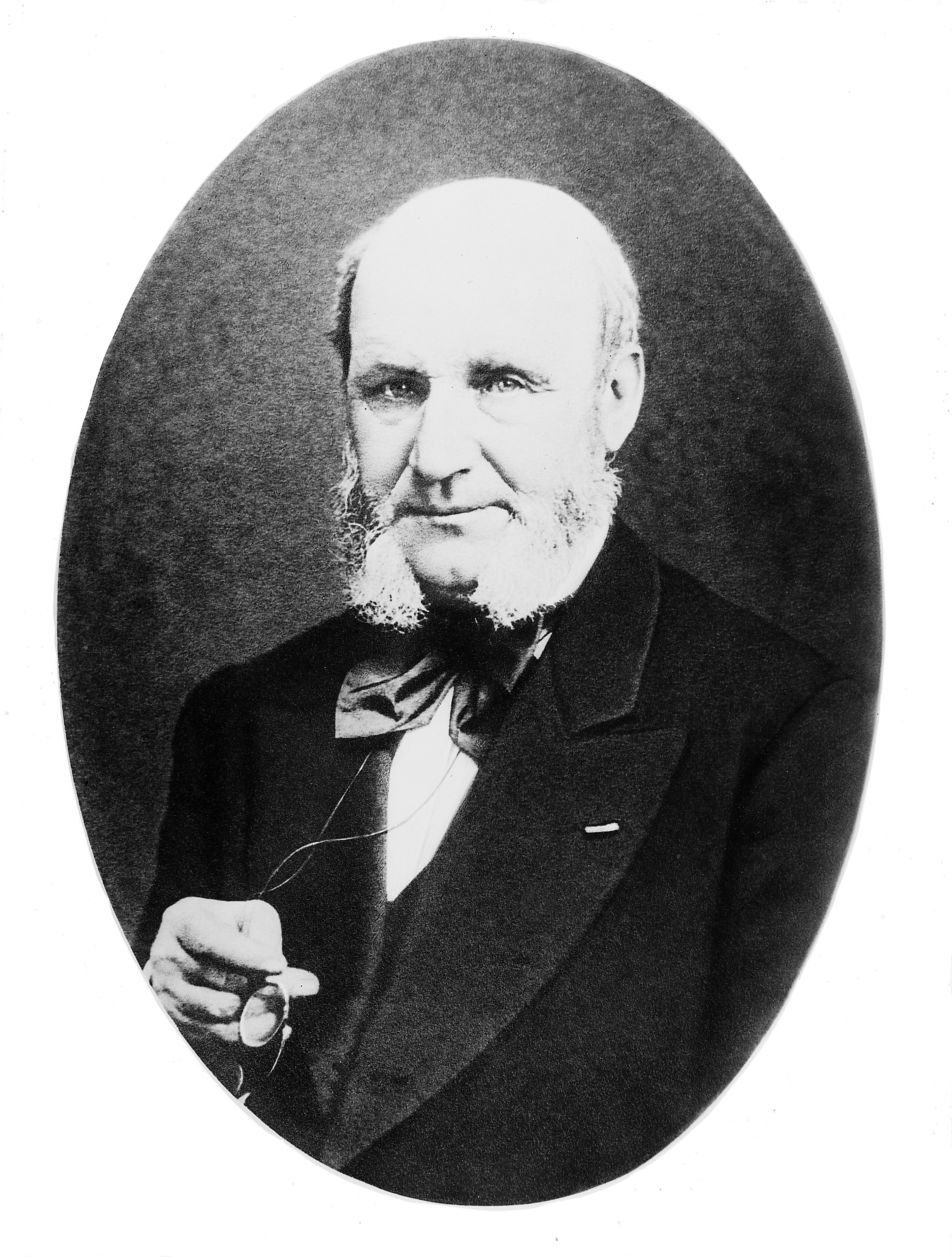
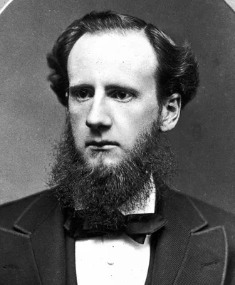
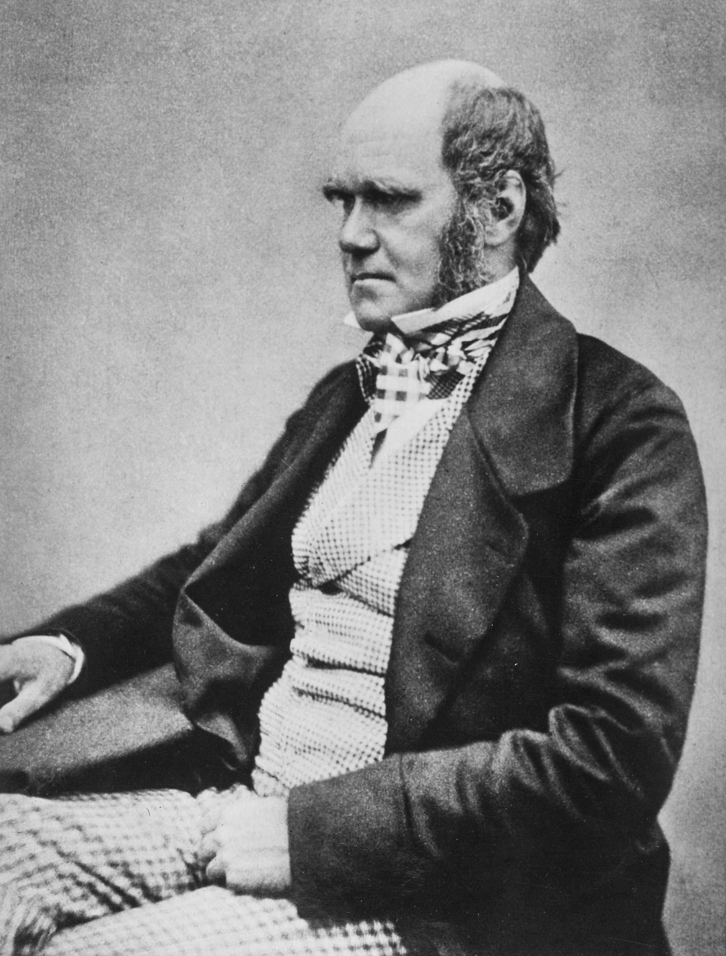
1844 –1845 DONNÉ, Alfred François (1801 – 1878); FOUCAULT, Jean Bernard Léon (1819 – 1868)
Cours de microscopie. 1 vol. and atlas.
Donne's and Foucault's work was the first biomedical textbook to be illustrated with images made from photomicrographs, in this case daguerreotypes of blood cells. Among its noteworthy images are the first microphotog…
GMN 267.1 · https://historyofmedicine.com/id/8743
1855 DUCHENNE DE BOULOGNE, Guillaume Benjamin Armand (1806 – 1875)
De l’électrisation localisée et de son application à la physiologie, à la pathologie, et à la thérapeutique.
Duchenne classified the electrophysiology of the entire muscular system and summed up his findings in the above work. The application of his results to pathological conditions marks him as the founder of electrotherap…
GMN 1995 · https://historyofmedicine.com/id/1848
1861 –1867 RÜDINGER, Nikolaus (1832 – 1896)
Atlas des peripherischen Nervensystems des menschlichen Körpers / Atlas du système nerveux périphérique du corps humain. Mit einem Vorwort von Prof. Dr. Th. W. L. Bischoff, nach der Natur photographirt von Joseph Alb. 10 parts.
Issued in 10 parts in folio (510 x 360 mm). Includes 46 large-format original photographs mounted on sheets of cardboard.
GMN 14159 · https://historyofmedicine.com/id/16474
1862 DUCHENNE DE BOULOGNE, Guillaume Benjamin Armand (1806 – 1875)
Mécanisme de la physionomie humaine, ou analyse électro-physiologique de l’expression des passions applicable à la pratique des arts plastiques. Premier fascicule. [All published]. 1 volume of text plus atlas of photographs by Duchenne.
Duchenne studied the mechanism of facial expression during emotion; his atlas of photographs was the first medical book illustrated with photographs of living subjects. Darwin reproduced a number of his photographs in…
GMN 4973 · https://historyofmedicine.com/id/6392
1864 DEAN, John (1831 – 1888)
The gray substance of the medulla oblongata and trapezium.
The first American medical book illustrated with photomechanically reproduced plates. Oliver Wendell Holmes praised the book for its remarkable photomicrographs, which may be the first published of brain cross-section…
GMN 7331 · https://historyofmedicine.com/id/9502
1865 SQUIRE, Alexander John Balmanno (1836 – 1908)
Photographs (coloured from life) of the diseases of the skin.
The first dermatologic publication illustrated with photographs.
GMN 6873 · https://historyofmedicine.com/id/9037
1868 HARDY, Alfred Louis Philippe (1811 – 1893); MONTMÉJA, Pierre Michel Arthur de (1841 – 1910)
Clinique photographique de l'hôpital Saint-Louis.
The first French dermatologic book illustrated with photographs. Originally issued in fascicules, 1867-68. The first edition includes 49 or 50 albumin photographs; second edition, entitled Clinique photographique des …
GMN 6872 · https://historyofmedicine.com/id/9036
1871 MONTMÉJA, Pierre Michel Arthur de (1841 – 1910); MEYER, Édouard (1838 – 1902)
Traité des opérations qui se pratiquent sur l'oeil.
Published in fascicules beginning in 1870. Includes 22 original mounted albumen photographs by Montméja illustrating chapters on cataract, iridectomy, strabismus, eyelids, and lacrymal passages. There are also 190 woo…
GMN 10123 · https://historyofmedicine.com/id/12312
1871 –1872 DUHRING, Louis Adolphus (1845 – 1913); MAURY, Francis Fontaine (1840 – 1879)
Photographic review of medicine and surgery. A bi-monthly illustration of interesting cases, accompanied by notes. Edited by F.F. Maury [and] L.A. Duhring. Vols. 1 & 2 (All published).
The leading 19th century American publication of artistic medical photography. Each of the two volumes includes 24 mounted photographs. The photographs ilustrate cases of unusual and extreme disease, such as gross def…
GMN 10585 · https://historyofmedicine.com/id/12778
1872 DARWIN, Charles Robert (1809 – 1882)
The expression of the emotions in man and animals.
Darwin examined the causes, physiological and psychological, of all the fundamental emotions in man and animals. He concluded that “the chief expressive actions exhibited by man and by the lower animals are now innate…
GMN 4975 · https://historyofmedicine.com/id/6395
1873 LUYS, Jules Bernard (1828 – 1897)
Iconographie photographique des centres nerveux.
Contains 70 photographs of brain sections taken by Luys himself, with 64 lithographed schemas based on his drawings. Luys undertook this work when the evidence of his lithographs published in 1865 (No. 4012) was dispu…
GMN 1406.01 · https://historyofmedicine.com/id/2066
1873 RIVIÈRE, Émile (1835 – 1922)
Découverte d’un squelette humain de l’époque paléolithique dans les cavernes des Baoussé-Roussé dites grottes de Menton.
In March 1872 Rivière discovered an entire fossil human skeleton in a cave at Menton, in the south of France near the Italian border. The skeleton, later known as “Menton man,” closely resembles the Cro-Magnon remains…
GMN 11333 · https://historyofmedicine.com/id/13531
1876 –1880 CHARCOT, Jean Martin (1825 – 1893); BOURNEVILLE, Désiré-Magloire (1840 – 1909); REGNARD, Paul-Marie-Léon (1850 – 1927)
Iconographie photographique de la Salpêtrière. Service de M. Charcot. 3 vols.
A photographic atlas devoted to cases of hysteria and epilepsy, with case histories; the third volume includes discussions of hypnotism, somnambulism and magnetism. Bourneville was Charcot’s assistant at the Salpêtriè…
GMN 4558.1 · https://historyofmedicine.com/id/8918
1876 DAGONET, Henri (1823 – 1902)
Nouveau traité élémentaire et pratique des maladies mentales. Suivi de considérations pratiques sur l'administration des asiles d'aliénés.
The first psychiatric textbook to utilize photographic illustrations of patients. This was the second edition of Dagonet's Traité élémentaire et pratique des maladies mentales (1862). The second edition included 8 Woo…
GMN 13103 · https://historyofmedicine.com/id/15356
1877 KOCH, Robert Heinrich Hermann (1843 – 1910)
Verfahrungen zur Untersuchung, zum Conserviren und Photographiren der Bacterien.
Koch greatly improved staining methods; he laid the foundations of the technical procedures employed in bacteriology today. In the above paper he described his method of slide preparation, making films of bacteria on …
GMN 2488 · https://historyofmedicine.com/id/3111
1877 SAYRE, Lewis Albert (1820 – 1900)
Spinal disease and spinal curvature, their treatment by suspension and the use of the plaster of Paris bandage.
Sayre’s monograph on his methods of treating tuberculosis of the spine and scoliosis is the first American surgical textbook to contain actual mounted photographs, some of which are remarkable for their artistic quali…
GMN 4344.1 · https://historyofmedicine.com/id/5717
1877 –1879 DUPUYTREN, Guillaume, Le Baron (1777 – 1835); HOUEL, Charles-Nicolas (1815 – 1881)
Catalogue des pièces du Musée Dupuytren, publié sous les auspices de la Faculté de Médecine de Paris. 5 vols. text plus 4 vols. atlas.
Detailed catalogue, illustrated by photography, of the Musée Dupuytren, a museum of gross pathology. When this catalogue was published the museum contained about 6000 items.
GMN 11147 · https://historyofmedicine.com/id/13344
1878 MAREY, Étienne Jules (1830 – 1904)
La méthode graphique dans les sciences expérimentales et particulièrement en physiologie et en médecine.
Marey pioneered the use of graphical recording in the experimental sciences, using instruments (many of his own invention) to capture and display data impossible to observe with the senses alone, and to record the pro…
GMN 7294 · https://historyofmedicine.com/id/9464
1880 FOX, George Henry (1846 – 1937)
Photographic illustrations of skin diseases.
Fox, who was Professor of Dermatology in New York, produced a valuable atlas of skin diseases.
GMN 3996 · https://historyofmedicine.com/id/5054
1880 GINÉ Y PARTAGÁS, Juan (1836 – 1903)
Tratado clinico iconografico de dermatologia quirurgica.
The first medical text in Spanish to use photographs as illustrations. Includes 17 lithographed plates (12 chromolithographs) and 3 original photographs (2 hand-colored). Giné y Partagás was a surgeon, dermatologist, …
GMN 6874 · https://historyofmedicine.com/id/9038
1882 FRENCH, Thomas Rushmore (1849 – 1929)
On photographing the larynx.
French was the first to obtain good photographs of the larynx.
GMN 3290 · https://historyofmedicine.com/id/4183
1883 –1884 BURDON-SANDERSON, Sir John Scott (1828 – 1905); PAGE, Frederick James Montague (1848 – 1907)
On the electrical phemomena of the excitatory process in the heart of the frog and of the tortoise, as investigated photographically.
See No. 824. This paper contains several tracings of the heart's electrical activity recorded with a capillary electrometer, the earliest graphic recorder of bioelectric signals. These were the "first undistorted trac…
GMN 831 · https://historyofmedicine.com/id/318
1884 FRENCH, Thomas Rushmore (1849 – 1929)
On a perfected method of photographing the larynx.
By means of a special camera of his own invention French improved the method of photographing the larynx.
GMN 3291 · https://historyofmedicine.com/id/4185
1885 DALTON, John Call (1825 – 1889)
Topographical anatomy of the brain. 3 vols.
The most outstanding American neurological atlas of the nineteenth century and one of the best American photographically illustrated medical books of the period. The atlas reproduces the specimens, which Dalton prepar…
GMN 7332 · https://historyofmedicine.com/id/9503
1886 –1890 MANSUROV, Nikolai Porfir'evich (1834 – 1892)
Klinicheskii sbornik po dermatologii i sifilologii. 4 vols.
Mansurov was the first dermatologist in Russia, and one of the first physicians to use photography systematically in medical illustration. This was a periodical, illustrated with original photographs, of which Manusro…
GMN 7744 · https://historyofmedicine.com/id/9916
1887 CROOKSHANK, Edgar March (1858 – 1928)
Photography of bacteria. Illustrated with eight-six photographs reproduced in autotype.
The first book entirely devoted to the photography of bacteria. Digital facsimile from Google Books at this link.
GMN 10596 · https://historyofmedicine.com/id/12789
1888 –1918 CHARCOT, Jean Martin (1825 – 1893); RICHER, Paul Marie Louis Pierre (1849 – 1933); GILLES DE LA TOURETTE, Georges Albert Édouard Brutus (1857 – 1904); LONDE, Albert (1858 – 1917)
NOUVELLE Iconographie de la Salpêtriere. 28 vols.
Henry Meige, Richer, and other pupils of Charcot published many valuable studies of the constitutional aspects of nervous diseases in the above work, a series unique in the history of medicine and of great value for t…
GMN 4575 · https://historyofmedicine.com/id/8973
1889 HUMPHRY, Sir George Murray (1820 – 1896)
Old age: The results of information received respecting nearly nine hundred persons who had attained the age of eighty years, including seventy-four centenarians.
Analysis, illustrated with Woodburytype photographs, of data collected by the "Collective Investigation Committee" of the British Medical Association. Note that in the 1880s attaining the age of 80 was considered wort…
GMN 10618 · https://historyofmedicine.com/id/12811
1890 BURDON-SANDERSON, Sir John Scott (1828 – 1905)
Photographic determination of the time-relations of the changes which take place in muscle during the period of so-called ‘latent stimulation.’
Measurement by means of photography, of the speed of the nervous impulse.
GMN 1284 · https://historyofmedicine.com/id/2307
1891 –1893 HIRST, Barton Cooke (1861 – 1935); PIERSOL, George Arthur (1856 – 1924)
Human monstrosities. 4 vols.
The first large work on the subject illustrated primarily by photographs of specimens, and one of the last to use to the term "monster" as a medical descriptor. Digital facsimile of the 4 volumes from the Internet Arc…
GMN 534.68 · https://historyofmedicine.com/id/126
1891 PIFFARD, Henry Granger (1842 – 1910); FULLER, Robert M.
A practical treatise on diseases of the skin
The first systematic treatise on dermatology published in America. Piffard was a pioneer of "indoor" magnesium flashlight photography, and took most of the 50 photographs reproduced in this book by this technique. Dig…
GMN 10598 · https://historyofmedicine.com/id/12791
1893 LONDE, Albert (1858 – 1917)
La photographie médicale: Application aux sciences médicales et physiologiques.
The first book on medical photography. Digital facsimile from http://jubilotheque.upmc.fr at this link.
GMN 8094 · https://historyofmedicine.com/id/10270
1894 MAREY, Étienne Jules (1830 – 1904)
Le mouvement.
Marey, like Muybridge (No. 650-51), was a pioneer in the use of serial pictures as a method of studying the mechanics of locomotion. English translation, 1895.
GMN 643 · https://historyofmedicine.com/id/522
1894 NITZE, Maximilian Carl-Friedrich (1848 – 1906)
Kystophotographischer Atlas.
Digital facsimile from the Internet Archive at this link.
GMN 8970 · https://historyofmedicine.com/id/11149
1894 CURSCHMANN, Heinrich (1846 – 1910)
Klinische Abbildungen: Sammlung von Darstellungen der Veränderung der äusseren Körperform bei inneren Krankheiten.
Includes 57 fine heliogravure reproductions of artistic photographs of disease, including numerous congenital deformities. Digital facsimile from Google Books at this link.
GMN 10595 · https://historyofmedicine.com/id/12788
1895 WILSON, Edmund Beecher (1856 – 1939); LEAMING, Edward (1861 – 1916)
An atlas of the fertilization and karyokinesis of the ovum.
The first atlas of photomicrographs showing fertilization and cellular development during mitosis. The photomicrographs were taken by Leaming. Digital facsimile from the Internet Archive at this link.
GMN 8653 · https://historyofmedicine.com/id/10830
1896 LEAMING, Edward (1861 – 1916); STARR, Moses Allen (1854 – 1932)
Atlas of nerve cells. With the cooperation of Oliver S. Strong and Edward Leaming.
Photomicrographs by Leaming. Digital facsimile from the Biodiversity Heritage Library at this link.
GMN 9639 · https://historyofmedicine.com/id/11826
1896 EDER, Josef Maria (1855 – 1944); VALENTA, Eduard (1857 – 1937)
Versuche über Photographie mittelst der Röntgen’schen Strahlen.
"Eder was the director of an institute for graphic processes and the author of an early history of photography. With the photochemist Valenta, he produced a portfolio in January 1896, less than a month after Wilhelm C…
GMN 10607 · https://historyofmedicine.com/id/12800
1896 KÖNIG, Carl Georg Walter (1859 – 1936)
14 Photographien mit Röntgen-strahlen aufgenommen im physikaloschen Verein zu Frankfurt A. M.
This collection of x-ray photographs published within a few months of Röntgen's discovery includes applications in archaeology and anthropology (x-rays of mummies) and forensic medicine (for the investigation of gunsh…
GMN 10608 · https://historyofmedicine.com/id/12801
1897 FOX, George Henry (1846 – 1937)
Skin diseases of children. With twelve photogravure and chromographic plates, and sixty illustrations in the text.
Digital facsimile from the Internet Archive at this link.
GMN 10597 · https://historyofmedicine.com/id/12790
1899 MUYBRIDGE, Eadweard [JAMES EDWARD MUGGERIDGE] (1830 – 1904)
Animals in motion.
GMN 650 · https://historyofmedicine.com/id/528
1901 MUYBRIDGE, Eadweard [JAMES EDWARD MUGGERIDGE] (1830 – 1904)
The human figure in motion. An electro-photographic investigation of consecutive phases of muscular actions.
Muybridge, an Englishman, made exhaustive photographic investigations of consecutive animal movements while he was in America. More than 100,000 photographs were embodied in his Animal locomotion. An electro-photograp…
GMN 651 · https://historyofmedicine.com/id/553
1905 –1906 WATERSTON, David
The Edinburgh stereoscopic atlas of Anatomy. Edited by David Waterston. 5 vols.
The first large scale application of stereoscopic photography to anatomy. The 5 parts each contain 50 stereo cards on which are pasted original stereo photographs and corresponding printed descriptive text. Digital fa…
GMN 10599 · https://historyofmedicine.com/id/12792
1908 BEDIN, Alphonse (1878 – )
La photographie au laboratoire de médecine légale de l'Université de Nancy.
iIlustrated with 77 photographs, Bedin's work describes the methods and practices of forensic photography developed at Nancy between 1905 and 1908. The work is divided into two parts: The first part contains four chap…
GMN 12898 · https://historyofmedicine.com/id/15145
1913 GLÉNARD, Roger (1880 – 1964)
Le mouvement de l’intestin en circulation artificielle (chez les vertébrés). Thèses présentées a la Faculté des sciences de Paris.
Cinematographic studies of the movements of the intestines in animals. Digital facsimile from the Internet Archive at this link.
GMN 1031 · https://historyofmedicine.com/id/1000
1929 PORGES, Otto (1879 – 1968)
Ueber Gastrophotographie.
Introduction of gastrophotography.
GMN 3548 · https://historyofmedicine.com/id/4059
1933 VAN DE VELDE, Theodoor Hendrik (1873 – 1937)
Sex efficiency through exercises. Special physical culture for women, with 480 cinematographic and 54 full-page illustrations.
Includes a series of 12 "cinematographic" films on 480 flicker cards. In all the photographs frontal nudity of the female model is covered by clothing, though her buttocks are exposed in some views. Having previously …
GMN 13200 · https://historyofmedicine.com/id/15461
1934 BERNAL, John Desmond (1901 – 1971); HODGKIN, Dorothy Crowfoot (1910 – 1994)
X-Ray photographs of crystalline pepsin.
Bernal and Hodgkin took the first X-ray photograph of a protein structure—crystalline pepsin. They showed that crystals of pepsin give an X-ray diffaction pattern, beginning protein crystallography. This may also be t…
GMN 6923 · https://historyofmedicine.com/id/9088
1934 GESELL, Arnold Lucius (1880 – 1961)
An atlas of infant behavior: A systematic delineation of the forms and early growth of human behavior patterns... illustrated with 3,200 action photographs. Vol. l: Normative series, in collaboration with Helen Thompson and Catherine S. Amatruda. Selected bibliographies (p. 45). Vol. 2: Naturalistic series, in collaboration with Alice V. Keliher, Frances L. Ilg, and Jessie J. Carlson. (2 vols.)
Gesell, who originated the Child Study Center at Yale University, was the founder of the study of child development in the United States. He is best known for his groundbreaking studies of normal child development: be…
GMN 10609 · https://historyofmedicine.com/id/12802
1942 LURIA, Salvador Edward (1912 – 1991); ANDERSON, Thomas Foxen (1911 – 1991)
The identification and characterization of bacteriophages with the electron microscope.
First identification, observation under the electron microscope, and reproduction of photomicrographs of bacteriophages taken through the electron microscope. Digital facsimile, but with poor reproductions of the two …
GMN 14004 · https://historyofmedicine.com/id/16309