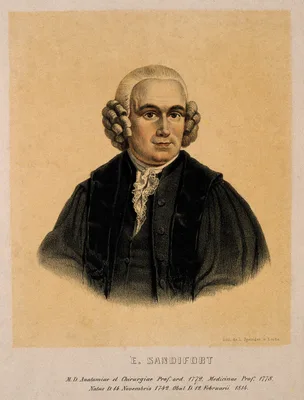
1777 –1781 SANDIFORT, Eduard (1742 – 1814)
Observationes anatomicae-pathologicae. 4 vols.
Sandifort’s beautifully illustrated work on pathological anatomy included records of ulcerative aortic endocarditis, renal calculi, hemias, bony ankyloses, and congenital abnormalities. A good account of the “tetralog…
GMN 2278 · https://historyofmedicine.com/id/2893
1787 DENMAN, Thomas (1733 – 1815)
A collection of engravings, tending to illustrate the generation and parturition of animals, and of the human species.
An idiosyncratic collection of rarely reproduced images with explanatory commentaries in English and French, concerning reproduction and obstetric complications in animals and humans. Topics include: The Funis of a nu…
GMN 12163 · https://historyofmedicine.com/id/14373
1793 –1835 SANDIFORT, Eduard (1742 – 1814); SANDIFORT, Gerard (1779 – 1848)
Museum anatomicum academiae Lugduno-Batavae. 4 vols.
This work, complete in over 1000 pages, with hundreds of full page plates, was begun by Eduard Sandifort and completed 42 years later by his son Gerard. Many of the plates illustrate diseases of bone. Digital facsimil…
GMN 11312 · https://historyofmedicine.com/id/13510
1799 –1803 BAILLIE, Matthew (1761 – 1823)
A series of engravings, accompanied with explanations, which are intended to illustrate the morbid anatomy of some of the most important parts of the human body.
The first systematic atlas of pathology. This work was intended to illustrate No. 2281, but, with its extensive descriptive text for each plate, it may be appreciated separately. The black & white engravings were prep…
GMN 2282 · https://historyofmedicine.com/id/2899
1817 ALIBERT, Jean Louis Marc (1768 – 1837)
Nosologie naturelle, ou les maladies du corps humain distribuées par familles. Tome premier. (All published.)
This unfinished work with 22 spectacular plates included some full body images of patients, and unlike other illustrated works by Alibert, it concerned more than skin diseases. Vol. 2 was never published but a second,…
GMN 12746 · https://historyofmedicine.com/id/14992
1826 HOOPER, Robert (1773 – 1835)
The morbid anatomy of the human brain.
Based on over 4000 autopsies performed over 30 years, and illustrated with fine hand-colored plates.
GMN 2284.1 · https://historyofmedicine.com/id/2904
1827 –1831 BRIGHT, Richard (1789 – 1858)
Reports of medical cases, selected with a view of illustrating the symptoms and cure of diseases by a reference to morbid anatomy. 2 vols. in 3.
Beside's Bright's classic description of chronic non-suppurative nephritis, known eponymically as “Bright’s disease”, the Reports contain numerous other outstanding contributions to general pathology, neuropathology, …
GMN 2285 · https://historyofmedicine.com/id/2906
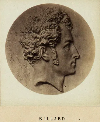
1828 BILLARD, Charles-Michel (1800 – 1832)
Traité des maladies des enfans nouveau-nés et à la mamelle. 1 vol. and atlas.
The first significant work on the pathological anatomy of infants. Billard performed several hundred autopsies on infants and children and correlated the data obtained with clinical observations he had made. This pion…
GMN 2285.1 · https://historyofmedicine.com/id/2908
1828 ANNESLEY, Sir James (1780 – 1847)
Researches into the causes, nature and treatment of the more prevalent diseases of India, and of warm climates generally. Illustrated with cases, post mortem examinations, and numerous coloured engravings of morbid structures. 2 vols.
A landmark in geographical pathology, superbly illustrated. Annesley’s cases, collected over many years’ service throughout India, represented the most complete treatment of diseases on the sub-continent to date. Digi…
GMN 1776.1 · https://historyofmedicine.com/id/3342
1829 –1842 CRUVEILHIER, Jean (1791 – 1874)
Anatomie pathologique du corps humain. 2 vols.
The fine hand-colored lithographs of gross pathology make this one of the greatest works of its kind. Cruveilhier, first Professor of Pathological Anatomy in Paris, gave the first description of multiple sclerosis (in…
GMN 2286 · https://historyofmedicine.com/id/2912
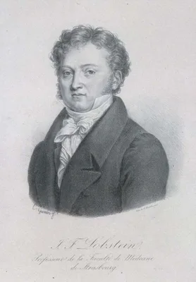
1829 –1833 LOBSTEIN, Jean Georges Chrétien Frédéric Martin (1777 – 1835)
Traité d’anatomie pathologique. 2 vols. and atlas.
Includes a historical review of the subject from the time of the Ancient Egyptians to Corvisart, and a summary of the advances in pathology during the preceding 50 years. Vol. 2, pp. 553-600 deals with diseases of the…
GMN 2288 · https://historyofmedicine.com/id/2915
1832 –1862 ALBERS, Johann Friedrich Hermann (1805 – 1867)
Atlas der pathologischen Anatomie für praktische Aerzte. 4 parts in 1. With: Erläuterung zu dem Atlasse der pathologischen Anatomie für praktischen Aertze. 4 vols. in 5.
Albers' atlas of pathology, with 257 lithographed plates (93 hand-colored) covers diseases of the brain and spinal cord (Part 1), the throat (Part 2), the thorax (Part 3) and the abdomen (part 4); the five text volume…
GMN 14348 · https://historyofmedicine.com/id/16678
1834 HOPE, James (1801 – 1841)
Principles and illustrations of morbid anatomy.
Hope left a fine pathological atlas with brilliantly hand-coloured lithographs from his own drawings. While the book does not equal the atlases of Cruveilhier and Carswell, it is important as being a great stimulus to…
GMN 2289 · https://historyofmedicine.com/id/2916
1834 MORTON, Samuel George (1799 – 1851)
Illustrations of pulmonary consumption.
Morton published an important collection of illustrations delineating pulmonary tuberculosis which epitomized the knowledge of his time. It was also the first book on the subject published in the United States. Digita…
GMN 3222 · https://historyofmedicine.com/id/5011
1834 GRANVILLE, Augustus Bozzi (1783 – 1872)
Graphic illustrations of abortion and the diseases of menstruation. Consisting of twelve plates from drawings engraved on stone, and coloured by Mr. J. Perry, and two copperplates from the Philosophical transactions, coloured by the same artist. The whole representing forty-five specimens of aborted ova and adventitious productions of the uterus, with preliminary observations, explanations of the figures, and remarks anatomical and physiological.
Concerns miscarriage and aberrant gestation rather than removal or expulsion of embryo or fetus. Digital facsimile from wellcomecollection.org at this link.
GMN 14017 · https://historyofmedicine.com/id/16324
1838 CARSWELL, Sir Robert (1793 – 1857)
Illustrations of the elementary forms of disease.
Carswell was Professor of Morbid Anatomy at University College, London, and one of the leading English pathologists of his day. A fine artist, he personally painted 2,000 water-colours of pathological specimens. His g…
GMN 2291 · https://historyofmedicine.com/id/2920
1839 –1841 RAYER, Pierre François Olive (1793 – 1867)
Traité des maladies des reins. 3 vols, and atlas.
Rayer insisted on the exhaustive analysis of the urine as an aid to the diagnosis of lesions. He classified “albuminus nephritis” into six distinct forms and distinguished these from other forms of nephritis associate…
GMN 4208 · https://historyofmedicine.com/id/6240
1839 BAUMGÄRTNER, Karl Heinrich (1798 – 1886)
Physiognomice pathologica – Krankenphysiognomik. Text in quarto; atlas in folio with 72 hand-colored lithographs.
A second edition in octavo format with 80 small plates was published in 1842. Baumgärtner, a pupil of Friedrich Tiedemann and Leopold Gmelin at Heidelberg, taught that it was possible to make a correct diagnosis with …
GMN 10589 · https://historyofmedicine.com/id/12782
1841 SEWALL, Thomas (1786 – 1845)
The pathology of drunkeness, or the effects of alcoholic drinks with drawings of the drunkard's stomach.
Including four chromolithographed plates by J. H. Hall, Albany, N.Y., this is the earliest illustrated book published in the U.S. on the pathological effects of alcoholism. Digital facsimile from the U.S. National Lib…
GMN 13521 · https://historyofmedicine.com/id/15796
1847 DANIELSSEN, Daniel Cornelius (1815 – 1894); BOECK, Carl Wilhelm (1808 – 1875)
Om spedalskhed. Udgivet efter Foranstaltning af den Kongelige Norske Regjerings Department for det Indre. 1 vol. and atlas.
First modern description of leprosy (“Danielssen-Boeck disease’’). Danielssen, physician to the leprosy hospital at Bergen, was the founder of scientific leprology. The extremely rare Atlas consists of 24 plates and t…
GMN 2434 · https://historyofmedicine.com/id/2894
1848 –1851 TARDIEU, Auguste Ambroise (1818 – 1879); AUVERT, Alexandre Ivanovich ( – 1865)
Selecta praxis medico-chirurgicae quam Mosquae exercet Alexander Auvert, ...Typis et figuris expressa Parisiis, moderante Ambroise Tardieu.
Auvert was professor in Moscow. This folio atlas includes 120 folio plates of pathology subjects, drawn by Schtschegoleff in Moscow. The text is in Latin throughout. The drawings were engraved in Paris by Oudet, print…
GMN 7495 · https://historyofmedicine.com/id/9667
1851 RICORD, Philippe (1800 – 1889)
Traité complet des maladies vénériennes. Clinique iconographique de l'Hôpital des vénériens. Recueil d'observations, suivies de considérations pratiques sur les maladies qui ont été traitées dans cet hôpital.
Ricord's extensive work with veneral diseases at the Hopital du Midi includes 66 hand-colored ithographs illustrating a multitude of conditions at various levels of infection in both sexes. The illustrations were draw…
GMN 14130 · https://historyofmedicine.com/id/16441
1857 –1861 LEBERT, Hermann (1813 – 1878)
Traité d’anatomie pathologique générale et spéciale. 4 vols.
Lebert set out to cover both general and special pathology. The superb hand-colored folio-sized copperplate engravings of macro- and micropathology in this work are among the finest ever published.
GMN 2297.1 · https://historyofmedicine.com/id/2931
1873 –1875 GOWERS, Sir William Richard (1845 – 1915); PAGENSTECHER, Hermann (1844 – 1932); GENTH, Carl Philipp (1844 – 1904)
Atlas der pathologischen Anatomie des Augapfels. Atlas of the pathological anatomy of the eyeball
Text in German and English; Sir W. R. Gowers was responsible for the English translation. Digital facsimile from the Bayerische StaatsBibliothek at this link.
GMN 5910 · https://historyofmedicine.com/id/6787
1873 –1892 SANGALLI, Giacomo Francesco (1821 – 1897)
La scienza e la pratica della anatomia patologica.
Divided into six parts, as follows: Book I: Delle alterazioni di prima formazione (on teratology) Book II: Delle ipertrofie Book III: Delle atrofie Book IV: Dell’infiammazione e della mortificazione Book V: Dei tumori…
GMN 7473 · https://historyofmedicine.com/id/9645
1875 ROKITANSKY, Carl Freiherr von (1804 – 1878)
Die Defecte der Scheidewände des Herzens.
Rokitansky’s memoir on defects of the septum of the heart was his last work, and possibly his greatest. It represented 14 years’ study of the subject.
GMN 2778 · https://historyofmedicine.com/id/4739
1877 –1907 HUTCHINSON, Sir Jonathan (1828 – 1913); ET AL
An atlas of illustrations of pathology [and] An atlas of illustrations of clinical medicine, surgery and pathology ... a continuation of the "Atlas of Pathology." 30 fascicules numbered I - XXIV, XXIVbis, XXIVter, XXV, XXVbis, XXVI - XXVII.
Bay far the most elaborate publication published by The New Sydenham Society, issued in fascicules over 30 years and rarely found complete.
GMN 14349 · https://historyofmedicine.com/id/16679
1882 DELAFIELD, Francis A. (1841 – 1915)
Studies in pathological anatomy. Vol. 1. Plates I. - XCIII. (All Published).
Delafield's work includes striking microscopic illustrations as well as explanatory text. Digital facsimile from Google Books at this link.
GMN 11351 · https://historyofmedicine.com/id/13549
1885 ESMARCH, Johann Friedrich August von (1823 – 1908); KULENKAMPFF, Diedrich (1846 – )
Die elephantiastischen Formen. Eine umfassende Darstellung der angeborenen und erworbenen Elephantiasis sowie aller verwandten Leiden.
This atlas illustrates the various changes that occur under the collective term elephantiasis, including tumors of the blood and lymphatic vessels, fibromas, neuromas, papillomas. Goldschmid 258.
GMN 9544 · https://historyofmedicine.com/id/11730
1892 –1896 BRAMWELL, Sir Byrom (1847 – 1931)
Atlas of clinical medicine. 3 vols.
Published at the end of the 19th century, and employing the wide variety of illustration technologies then available, including color lithography, lithography, and photography, this work testifies to the breadth and d…
GMN 10621 · https://historyofmedicine.com/id/12814
1897 MEIGS, Arthur Vincent (1850 – 1912)
The origin of disease, especially of disease resulting from intrinsic as opposed to extrinsic causes. With chapters on diagnosis, prognosis and treatment. With one hundred and thirty-seven original illustrations.
The most valuable features of this work are the exceptionally fine and delicate histologic illustrations engraved on steel and printed on thick paper to eliminate show-through. Each image is faced with detailed explan…
GMN 11875 · https://historyofmedicine.com/id/14079
1925 GOLDSCHMID, Edgar (1881 – 1957)
Entwicklung und Bibliographie der pathologisch-anatomischen Abbildung.
Traces the development of pathological anatomical illustration, and includes a chronological bibliography of all important publications known to Goldschmid containing illustrations of pathological conditions, and an i…
GMN 2316 · https://historyofmedicine.com/id/2995
1995 SCHNALKE, Thomas (1958 – )
Diseases in wax: A history of the medical moulage.
GMN 7499 · https://historyofmedicine.com/id/9671
2017 BERTOLONI MELI, Domenico
Visualizing disease: The art and history of pathological illustrations.
GMN 9849 · https://historyofmedicine.com/id/12037