1538 ANONYMOUS
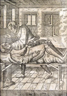
Ein newes hochnutzlichs Büchlin von erkantnus der Kranckheyten der Augen....
The first separate publication on diseases of the eye after Grassi (1474). This very rare anonymous work was intended, according to its title, to provide highly useful knowledge of the anatomy of the eye, and eye dise…
GMN 6932 · https://historyofmedicine.com/id/9097
1574 CARCANO LEONE, Giovanni Battista (1536 – 1606)
Anatomici libri II … In altero de musculis, palpebrarum atque oculorum motibus deservientibus, accurate disseritur.
First exact description of the lacrimal duct. Carcano gave the true position of the lacrimal gland and showed the route taken by the tears.
GMN 1479 · https://historyofmedicine.com/id/2462
1583 BARTISCH, Georg (1535 – 1606)
̓Oφθαλμоδоύλεια das ist, Augendienst.
In this treatise on ophthalmic surgery Bartisch, who limited his practice to ophthalmology and hernia repair, left the first extensively illustrated account of any surgical specialty. Bartisch was a skilful operator a…
GMN 5817 · https://historyofmedicine.com/id/7024
1587 ARANZI, Giulio Cesare [ARANZIO; ARANTIUS] (1530 – 1589)
De humano foetu liber tertio editus, ac recognitus. Eiusdem anatomicarum observationum liber: ac De tumoribus secundum locos affectos liber nunc primum editi.
First edition of Aranzi's Anatomicarum observationum published with the third edition of De humano foetu. In the Anatomicarum observationum Aranzi pointed out that the eye muscles arise from the margin of the optic ca…
GMN 7337 · https://historyofmedicine.com/id/9508
1666 MEIBOM, Heinrich (1638 – 1700)
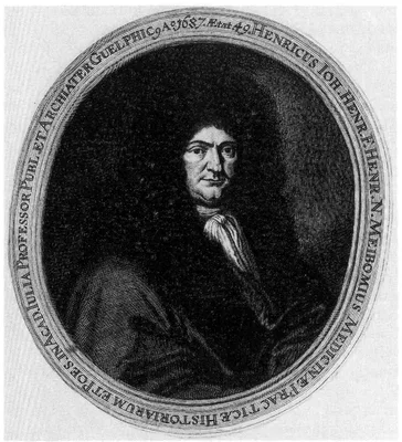
De vasis palpebrarum novis epistola.
Meibom described the conjunctival Meibomian glands, holocrine type exocrine glands along the rims of the eyelid inside the tarsal plate. They were, however, already known to Galen and were figured by Casserius in 1609.
GMN 1481 · https://historyofmedicine.com/id/2469
1676 BRIGGS, William (1642 – 1704)
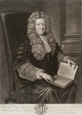
Ophthalmo-graphia; sive, oculi eiusque partium descriptio anatomica.
First English treatise on the anatomy of the eye. Briggs described the papilla of the optic disc and hypothesized that vibrations caused by rays of light striking fibers of the retina were conveyed to the papilla, and…
GMN 1481.2 · https://historyofmedicine.com/id/2476
1729 DUDDELL, Benedict (1695 – )
A treatise of the diseases of the horny coat of the eye, and the various kinds of cataracts.
“Descemet’s membrane” was first described by Duddell. Descemet described it in 1758; see No. 1484.1.
GMN 1482 · https://historyofmedicine.com/id/2489
1753 ZINN, Johann Gottfried (1727 – 1759)
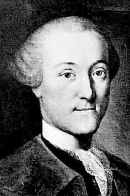
De ligamentis ciliaribus.
GMN 1483 · https://historyofmedicine.com/id/2494
1755 ZINN, Johann Gottfried (1727 – 1759)
Descriptio anatomica oculi humani.
The first complete study of the anatomy of the eye, including the first description of the “zonule of Zinn” and the “annulus of Zinn”.
GMN 1484 · https://historyofmedicine.com/id/2499
1758 DESCEMET, Jean (1732 – 1810)
An sola lens crystallina cataracte sedes?
“Descemet’s membrane”, the posterior membrane of the cornea; see No. 1482.
GMN 1484.1 · https://historyofmedicine.com/id/2509
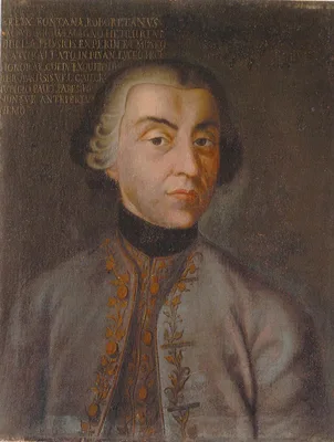
1767 FONTANA, Felice (1730 – 1805)
Ricerche fisiche sopra il veleno della vipera.
The starting point of modern investigations of serpent venoms and their antidotes. This work also includes Fontana’s description of the ciliary canal in the eye of an ox. This structure does not appear in the human ey…
GMN 2103 · https://historyofmedicine.com/id/2601
1801 SOEMMERRING, Samuel Thomas von (1755 – 1830)
Abbildungen des menschlichen Auges.
Soemmerring is best remembered for his fine anatomical illustrations, of which those devoted to the human eye are a good example. In 1791 he made important observations on the macula lutea: "De foramine centrali limbo…
GMN 1489 · https://historyofmedicine.com/id/2536
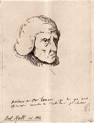
1806 TENON, Jacques René (1724 – 1816)
Observations anatomiques sur quelques parties de l’oeil et des paupières. IN: Mémoires et observations sur l’anatomie, la pathologie, et la chirurgerie, pp. 193-207.
Although Tenon did not discover the fibrous capsule and the interfascial space of the orbit, they are named after him.
GMN 1490 · https://historyofmedicine.com/id/2559
1819 JACOB, Arthur (1790 – 1874)
An account of a membrane in the eye, now first described.
“Jacob’s membrane”, the layer of the retina containing the rods and cones.
GMN 1491 · https://historyofmedicine.com/id/2560
1824 HORNER, William Edmonds (1793 – 1853)
Description of a small muscle of the internal commissure of the eyelids.
Homer described the tensor tarsi (Horner’s) muscle, supplying the lacrimal apparatus. It was first described by Du Verney in 1749
GMN 1494 · https://historyofmedicine.com/id/2570
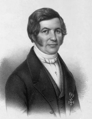
1830 SCHLEMM, Friedrich (1795 – 1858)
Arteriarum capitis superficialium icon nova.
Includes description of the “canal of Schlemm”, the circular canal at the junction of the cornea and the sclerotic.
GMN 1456.1 · https://historyofmedicine.com/id/2360
1832 FIELDING, George Hunsley (1801 – 1871)
On a new membrane in the eye.
“Fielding’s membrane”, the tapetum of the retina.
GMN 1496 · https://historyofmedicine.com/id/2574
1834 DALRYMPLE, John (1804 – 1852)
The anatomy of the human eye.
First English work on ocular anatomy.
GMN 1497 · https://historyofmedicine.com/id/2575
1844 BRUCH, Karl Wilhelm Ludwig (1819 – 1884)
Untersuchungen zur Kenntniss des körnigen Pigments der Wirbelthiere in physiologischer und pathologischer Hinsicht.
Includes a description of “Bruch’s membrane” of the choroid.
GMN 1501 · https://historyofmedicine.com/id/2579
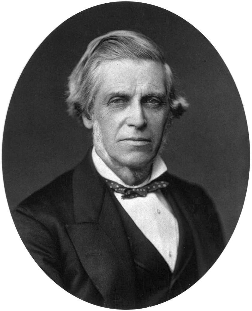
1849 BOWMAN, Sir William (1816 – 1892)
Lectures on the parts concerned in the operations on the eye, and on the structure of the retina.
Bowman did more than any other man to advance ophthalmic surgery in England. The above work is the first to include a sound description of the microscopical anatomy of the eye and the ciliary (“Bowman’s”) muscle. The …
GMN 1505 · https://historyofmedicine.com/id/2585
1851 MÜLLER, Heinrich (1820 – 1864)
Zur Histologie der Netzhaut.
Discovery of visual purple.
GMN 1506 · https://historyofmedicine.com/id/2589
1858 AMMON, Friedrich August von (1799 – 1861)
Die Entwicklungsgeschichte des menschlichen Auges.
"Ammon's Scleral Prominence - prominence of the postero-external aspect of the globe of the eye of the foetus which appears at the third month of foetal life." Jessie Dobson, eponyms, 2nd. Ed. (1962), p.13 . The artic…
GMN 13339 · https://historyofmedicine.com/id/15607
1866 SCHULTZE, Max Johann Sigismund (1825 – 1874)
Zur Anatomie und Physiologie der Retina.
One of the greatest of all histologists, Max Schultze is remembered by ophthalmologists for his monograph on the nerve-endings in the retina.
GMN 1512 · https://historyofmedicine.com/id/2605
1873 –1875 GOWERS, Sir William Richard (1845 – 1915); PAGENSTECHER, Hermann (1844 – 1932); GENTH, Carl Philipp (1844 – 1904)
Atlas der pathologischen Anatomie des Augapfels. Atlas of the pathological anatomy of the eyeball
Text in German and English; Sir W. R. Gowers was responsible for the English translation. Digital facsimile from the Bayerische StaatsBibliothek at this link.
GMN 5910 · https://historyofmedicine.com/id/6787
1876 SATTLER, Hubert (1844 – 1928)
Ueber den feineren Bau der Chorioidea des Menschen nebst Beiträgen zur pathologischen und vergleichenden Anatomie der Aderhaut.
“Sattler’s layer” of the choroid.
GMN 1518 · https://historyofmedicine.com/id/2622
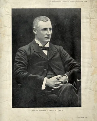
1885 LOCKWOOD, Charles Barrett (1856 – 1914)
The anatomy of the muscles, ligaments and fasciae of the orbit, including an account of the capsule of Tenon, the check ligaments of the recti, and the suspensory ligaments of the eye.
“Lockwood’s suspensory ligament” of the globe of the eye.
GMN 1520 · https://historyofmedicine.com/id/2626
1892 BOWMAN, Sir William (1816 – 1892); BURDON-SANDERSON, Sir John Scott (1828 – 1905); HULKE, John Whitaker (1830 – 1895)
The collected papers of Sir W. Bowman. Edited by J. Burdon-Sanderson and J. W. Hulke. 2 vols.
Digital facsimile from Google Books at this link.
GMN 12786 · https://historyofmedicine.com/id/15032
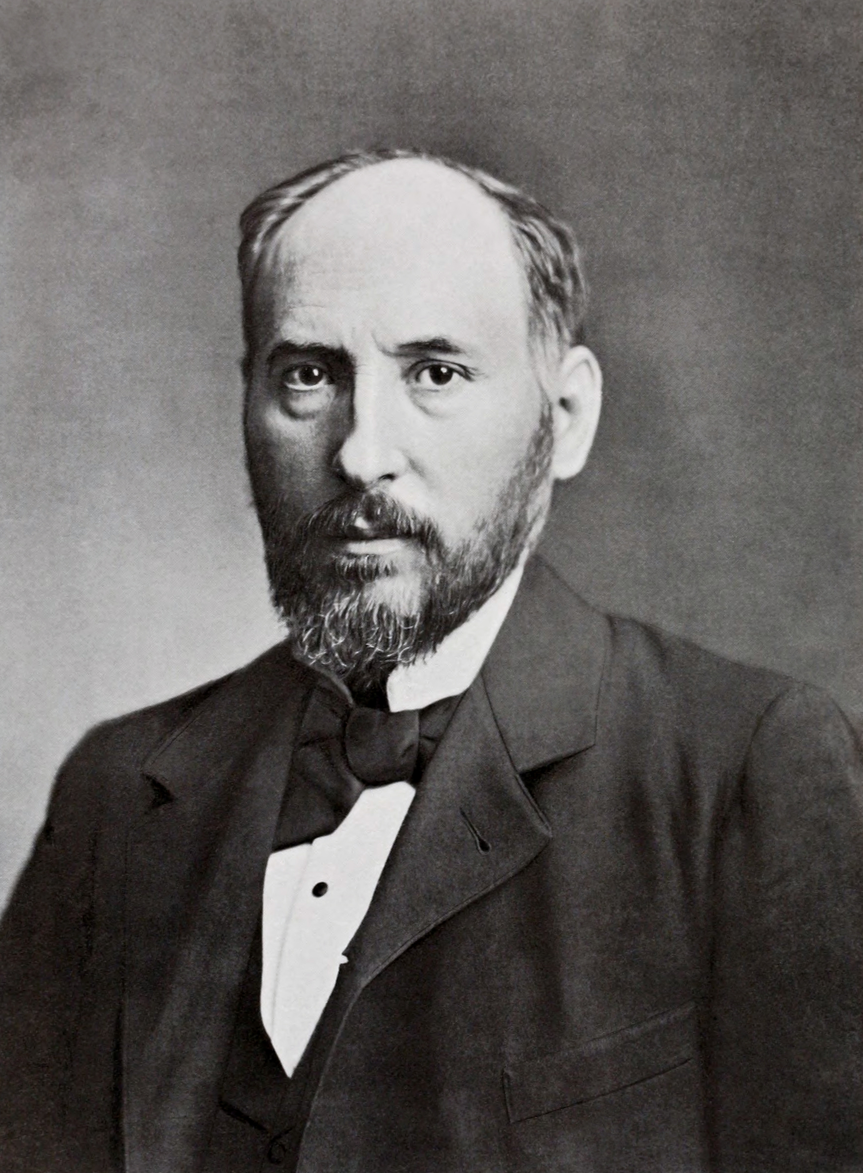
1894 CAJAL, Santiago Ramón y (1852 – 1934)
Die Retina der Wirbelthiere…In Verbindung mit dem Verfasser zusammengestellt, übersetzt und mit Einleitung versehen von Dr. Richard Greef.
Classic account of the vertebrate retina. First published in the Belgian review La Cellule, and later translated into German with extensive additions by Ramón y Cajal. Translated into English by Sylvia A. Thorpe and M…
GMN 1523 · https://historyofmedicine.com/id/3620
1921 VOGT, Alfred (1879 – 1943)
Atlas der Spaltlampenmikroskopie des lebenden Auges.
An important work on the biomicroscopy of the eye. Second ed. greatly revised and enlarged, vol. 1-2, Springer, 1930-31; vol. 3, Stuttgart, F. Enke, 1942; vol. 3 (English translation) Zurich, 1947. Second ed. reprinte…
GMN 1527 · https://historyofmedicine.com/id/2853
1928 MANN, Ida Caroline (1893 – 1983)
The development of the human eye.
The first monograph on the subject, illustrated with drawings by the author.
GMN 7392 · https://historyofmedicine.com/id/9564
1937 MANN, Ida Caroline (1893 – 1983)
Developmental abnormalities of the eye.
GMN 5987 · https://historyofmedicine.com/id/7073
1941 POLYAK, Stephen Lucian (1889 – 1955)
The anatomy and the histology of the retina in man, ape, and monkey.
A scholarly tour-de-force with a bibliography of over 700 references.
GMN 1533.1 · https://historyofmedicine.com/id/2860