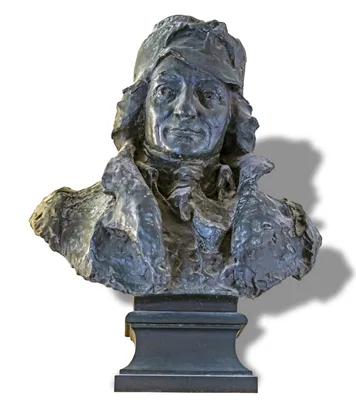
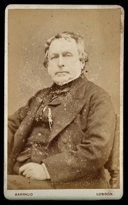
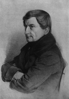
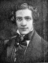
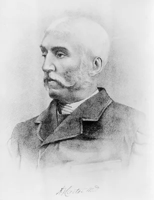
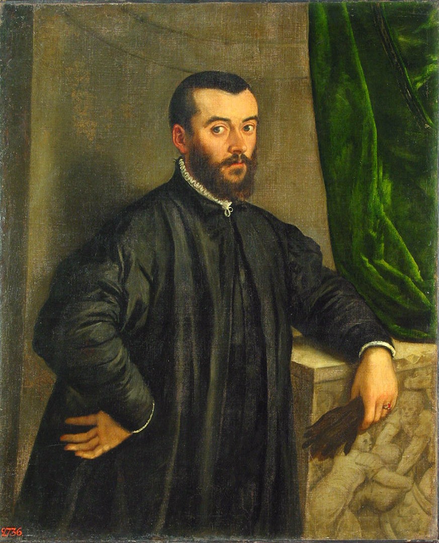
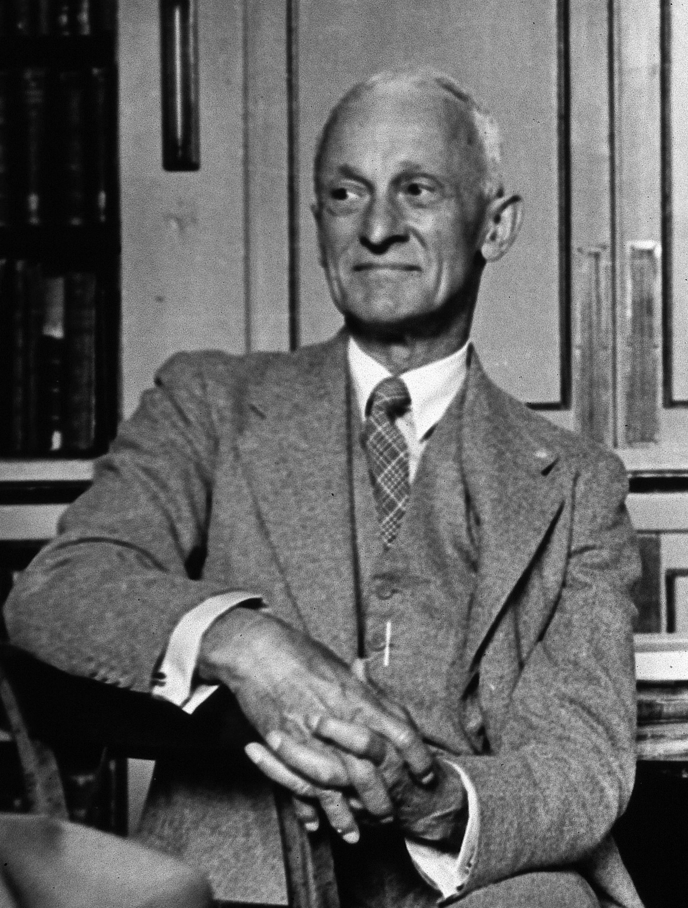
1741 PIETRO DA CORTONA, [Pietro BERRETINI] (1596 – 1669)
Tabulae anatomicae.
27 anatomical copperplates after drawings by the most influential painter of the Italian Baroque movement, who also excelled as an architect. The editor, Cajetano Petrioli, supplied the text and small numbered anatomi…
GMN 395.2 · https://historyofmedicine.com/id/695
1743 –1756 HALLER, Albrecht von (1708 – 1777)
Icones anatomicae. 8 pts.
Accurate and beautiful engravings of the diaphragm, uterus, ovaries, vagina, arteries, with explanatory observations. About fifty years after they were originally published the most visually spectacular versions of Ha…
GMN 397 · https://historyofmedicine.com/id/725
1745 GAUTIER D’AGOTY, Jacques Fabien (1716 – 1785); DU VERNEY, Guichard-Joseph (Guichard Joseph Duverney) (1648 – 1730)
Essai d’anatomie en tableaux imprimés, qui represent au naturel tous les muscles de la face, du col, de la tête, de la langue & du larinx. d'après les parties disséquées & préparées par Monsieur Duverney....comprenant hit grandes planches.
Remarkable for its striking mezzotints printed in color. Digital facsimile from the Internet Archive at this link.
GMN 398 · https://historyofmedicine.com/id/739
1748 GAUTIER D’AGOTY, Jacques Fabien (1716 – 1785); DU VERNEY, Guichard-Joseph (Guichard Joseph Duverney) (1648 – 1730)
Anatomie de la tête, en tableaux imprimés qui représentent au naturel le cerveau sous différentes coupes, la distribution des vaisseaux dans toutes les parties de la tête, les organes des sens et une partie de la névrologie, d'après les pièces disséquées et préparées par M. Duverney, en huit grandes planches dessinées, peintes, gravées et imprimées en couleur et grandeur naturelle....
Includes eight spectacular plates printed in color by Gautier d'Agoty with text by Duvereny. Digital facsimile from the Internet Archive at this link.
GMN 7491 · https://historyofmedicine.com/id/9663
1754 GAUTIER D’AGOTY, Jacques Fabien (1716 – 1785)
Anatomie generale des visceres, et de la nevrologie, angeologie et osteologie du corps humain, en figures, de couleurs et grandeurs naturelles dediée et présentée.
Includes 18 full-page color-printed mezzotints, 12 of the plates designed to fit together in threes to make four life-size human figures. Gautier credited Mertrud, the King's Surgeon with some of the anatomical work i…
GMN 7492 · https://historyofmedicine.com/id/9664
1756 JENTY, Charles Nicholas (1720 – 1770)
An essay on the demonstration of the human structure, half as large as nature, in four tables, from the pictures painted after dissections, for that purpose.
Four large mezzotint plates issued with an accompanying text.
GMN 7489 · https://historyofmedicine.com/id/9661
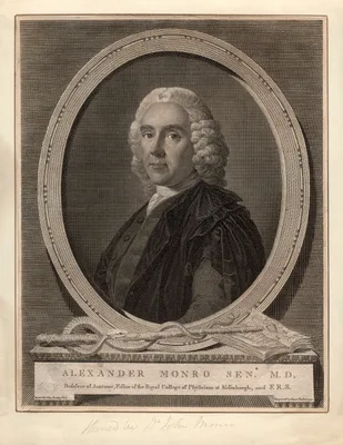
1759 MONRO, Alexander, Primus (1697 – 1767); SUE, Jean-Joseph (1710 – 1792)
Traité d'ostéologie. 2 vols.
Monro Primus' textbook on the anatomy of the bones was originally published in 1726 as an octavo volume without plates, and went through more than ten editions. The French translation, published in large folio, transl…
GMN 7006 · https://historyofmedicine.com/id/9172
1771 MOEHSEN, Johann Karl Wilhelm (1722 – 1795)
Verzeichnis einer Samlung von Bildnissen, gröstentheils berühmter Aerzte; so wohl in Kupferstichen, schwarzer Kunst und Holzschnitten, als auch in einigen Handzeichungen: diesem sin verschiedene Nachrichten und Unmerkungen Vorgesekt, die so wohl zur Geschichte der Arzenengelahrtheit; als vornehinsich zur Geschichte der Künste gehören.
“Very valuable reports on artistic anatomy and the history of anatomic illustration” (Choulant). Probably represents the first history of medical portraiture. Digital facsimile from Googe Books at this link.
GMN 6604.9 · https://historyofmedicine.com/id/8565
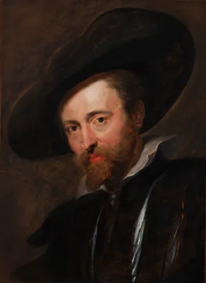
1773 RUBENS, Peter Paul (1577 – 1640)
Théorie de la figure humaine.
This work on the human figure, published more than 100 years after the death of Rubens, is one of a handful of anatomical treatises illustrated by an artist of the first magnitude.
GMN 155 · https://historyofmedicine.com/id/3164
1775 SANTORINI, Giovanni Domenico (1681 – 1737)
Septemdecim tabulae…
Santorini died before the completion of these anatomical plates which he intended to be his chef d’oeuvre. This elegantly printed volume is the only significant medical book printed by the celebrated Giambattista Bodo…
GMN 399.1 · https://historyofmedicine.com/id/748
1778 SOEMMERRING, Samuel Thomas von (1755 – 1830)
Dissertatio inauguralis anatomica de basi encephali et originibus nervorum cranio egredientium libri quinque.
The first accurate enumeration of the 12 cranial nerves, superseding that of Willis (No. 1378). Soemmerring is notable for his accuracy in anatomical illustration. This was his thesis. The same publisher issued an edi…
GMN 1383 · https://historyofmedicine.com/id/1808
1779 GAMELIN, Jacques (1738 – 1803)
Nouveau recueil d’ostéologie et de myologie…
“Without contest the most beautiful of all anatomies for the artist and one of the most remarkable books of its time” (Hahn & Dumaitre). The plates in this work are more fantastic than any other anatomy, suggesting th…
GMN 401.1 · https://historyofmedicine.com/id/781
1780 CATTANI, Antonio
Osteografia e miografia della testa, mani, e piedi del corpo umano in misura naturale con il catalogo de' nomi propri.
Very distinctive engravings with the captions included within the engraved surfaces. Printed and sold by the artist.
GMN 9547 · https://historyofmedicine.com/id/11733
1787 MASCAGNI, Paolo (1752 – 1815)
Vasorum lymphaticorum corporis humani historia et ichnographia.
Mascagni, Professor of Anatomy at Siena, made several discoveries regarding the lymphatics. His beautiful atlas contained 41 engravings of the lymphatics and gained him lasting fame. He had previously published a Prod…
GMN 1104 · https://historyofmedicine.com/id/1151
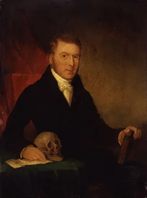
1794 BELL, John (1763 – 1820)
Engravings, explaining the anatomy of the bones, muscles and joints.
Bell’s atlas of the bones, muscles and joints was issued as a separate work a year after his text, The Anatomy of the Bones, Muscles, and Joints. Bell’s illustrations are some of the most striking in the entire litera…
GMN 9833 · https://historyofmedicine.com/id/12021
1797 SOEMMERRING, Samuel Thomas von (1755 – 1830)
Tabula sceleti feminini juncta descriptione.
Soemmerring was noted for his accuracy in anatomical illustration, and the above work is a fine example of his artistic sense. For it he selected the skeleton of a well-built girl of 20 years. Great care was taken in …
GMN 401 · https://historyofmedicine.com/id/775
1797 –1804 BELL, Sir Charles (1774 – 1842); BELL, John (1763 – 1820)
The anatomy of the human body. 4 vols.
“The first great textbook contributed by the British school to modern anatomy” (Russell, No. 461).
GMN 401.3 · https://historyofmedicine.com/id/797
1798 –1803 BELL, Sir Charles (1774 – 1842)
A system of dissections. 2 vols.
Published in 7 fascicules and appendix while Bell was still a student, this was Bell’s first independent venture as an author. The anatomical work of Charles Bell and his brother John was among the most significant in…
GMN 402 · https://historyofmedicine.com/id/813
1801 SOEMMERRING, Samuel Thomas von (1755 – 1830)
Abbildungen des menschlichen Auges.
Soemmerring is best remembered for his fine anatomical illustrations, of which those devoted to the human eye are a good example. In 1791 he made important observations on the macula lutea: "De foramine centrali limbo…
GMN 1489 · https://historyofmedicine.com/id/2536
1806 SOEMMERRING, Samuel Thomas von (1755 – 1830)
Abbildungen des menschlichen Hoerorganes
GMN 1554 · https://historyofmedicine.com/id/1173
1813 MITANI, Soshu (Koki) (1774 – 1823)
Kaitai hatsumo [Explanation of Human Anatomy]. 5 vols.
The first Japanese exposition of the whole system of human anatomy, based on original observation. The work includes the records of three dissections performed in Kyoto in 1783, 1798, and 1802. “A Japanese treatise on…
GMN 13214 · https://historyofmedicine.com/id/15477
1816 MASCAGNI, Paolo (1752 – 1815)
Anatomia per uso degli studiosi di scultura e pittura.
Mascagni's anatomy for artists and sculptors, edited for posthumous publication by Mascagni's literary execultor Francesco Antonmarchi. 15 hand-colored engraved plates after drawings by Antonio Serantoni (1780j-1837),…
GMN 7488 · https://historyofmedicine.com/id/9660
1818 RIEMER, Pieter de (1760 – 1831)
Afbeeldingen van de juiste plaatsing der inwendige deelen van het menschelijk ligchaam.
First anatomical illustrations of frozen sections. De Riemer appears to have been the first to freeze tissues in order to permit fine sectioning for the purposes of diagnosing diseased tissue. Digital facsimile from U…
GMN 408 · https://historyofmedicine.com/id/860
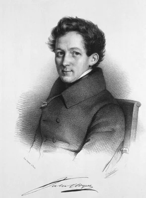
1821 –1831 CLOQUET, Jules Germain (1790 – 1883)
Anatomie de l’homme, ou descriptions et figures lithographiées de toutes les parties du corps humain. 5 vols.
The first anatomical atlas illustrated by lithography, containing 300 plates in folio format. This was one of the most elaborate of the lithographic “incunabula” produced by Charles Philibert de Lasteyrie, one of the …
GMN 409 · https://historyofmedicine.com/id/864
1822 –1832 MASCAGNI, Paolo (1752 – 1815)
Anatomia universa… 2 vols.
The largest of all medical books from the standpoint of format. The 44 life size engraved plates are reproduced in double elephant folio size measuring 950 x 635 mm., and include an almost incredible level of detail. …
GMN 409.1 · https://historyofmedicine.com/id/884
1822 –1826 LIZARS, John (1794 – 1860); LIZARS, William Home (1788 – 1859)
A system of anatomical plates of the human body. By John Lizars. Text in 8vo in 12 pts., plus folio atlas with engraved title and 101 plates engraved by William Home Lizars.
Lizars's System of anatomical plates was by far his most successful work, going through many editions. The text of the work was originally issued in 12 parts in octavo format, which were then bound together in book fo…
GMN 13508 · https://historyofmedicine.com/id/15783
1823 –1826 MASCAGNI, Paolo (1752 – 1815); ANTOMMARCHI, Francesco (1789 – 1838)
Planches anatomiques du corps humain executes d’après les dimensions naturelles. Double elephant folio atlas and small folio text.
Considering that it is among the rarest of all anatomies, and certainly the largest and probably the most spectacular, it is remarkable that two nearly identical editions of Mascagni’s posthumous life-size anatomy wer…
GMN 7242 · https://historyofmedicine.com/id/9411
1828 TUSON, Edward William (1802 – 1865)
Myology, illustrated by plates.
This large folio work contains 8 hand-colored lithographed plates by F. R. Say, each with multiple lift-up flaps. It is probably the largest format anatomical work ever published in English with lift-up flaps, with up…
GMN 10023 · https://historyofmedicine.com/id/12212
1830 HOME, Sir Everard (1756 – 1832)
A short tract on the formation of tumours, and the peculiarities that are met with in the structure of those that have become cancerous; with their mode of treatment.
Contains the first illustrations of microscopic sections of cancer; however, Home drew no worthwhile conclusion from his microscopic studies. Digital facsimile from the Internet Archive at this link.
GMN 2611 · https://historyofmedicine.com/id/3093
1831 –1854 BOURGERY, Jean Baptiste Marc (1797 – 1849); JACOB, Nicolas-Henri (1782 – 1871)
Traité complet de l'anatomie de l'homme, comprenant la médecine opératoire. 16 vols.
With over 2000 pages of text and 726 lithographed plates (incorporating 3604 individual figures), this work is the most comprehensive, and perhaps the most beautiful anatomical surgical atlas of the 19th century. It w…
GMN 7246 · https://historyofmedicine.com/id/9415
1833 FLAXMAN, John (1755 – 1826); LANDSEER, Sir Edwin Henry (1802 – 1873)
Anatomical studies of the bones and muscles, for the use of artists. From drawings by the late John Flaxman, Esq. R.A. Engraved by Henry Landseer. With two additional plates, and explanatory notes, by William Robertson.
Digital facsimile from digitalcollections.nypl.org at this link.
GMN 411 · https://historyofmedicine.com/id/912
1834 TUSON, Edward William (1802 – 1865)
The anatomy and surgery of inguinal and femoral hernia.
Published in the same large folio format as Tuson's Myology (1828), this was the largest work on hernia ever published with multiple hand-colored. lift-up flats on three plates.
GMN 10024 · https://historyofmedicine.com/id/12213
1836 –1842 QUAIN, Jones (1796 – 1865); WILSON, Sir William James Erasmus (1809 – 1884)
A series of anatomical plates. 5 vols.: The muscles of the human body. The vessels of the human body. The nerves of the human body. The viscera of the human body. The bones and ligaments of the human body.
The most ambitious 19th century English anatomy illustrated by lithography. Some copies were issued with hand-colored plates. The five volumes, containing a total of 201 plates, describe the muscles, blood vessels, ne…
GMN 13300 · https://historyofmedicine.com/id/15566
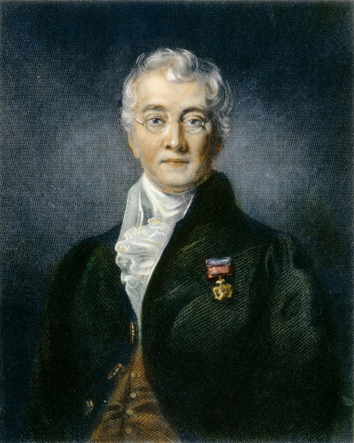
1852 –1859 PIROGOV, Nikolai Ivanovich (Pirogoff) (1810 – 1881)
Anatome topographica sectionibus per corpus humanum congelatum triplici directione ductis illustrata. 8 pts.
Pirogov was the greatest of Russian surgeons. He introduced the teaching of applied topographical anatomy in Russia. His atlas of 220 plates represents the first use on a grand scale of frozen sections in anatomical i…
GMN 416 · https://historyofmedicine.com/id/950
1852 CHOULANT, Johann Ludwig (1791 – 1861)
Geschichte und Bibliographie der anatomischen Abbildung.
In this classic work Choulant traced the evolution of anatomical illustration from the early schematic plates up to his own time, including a valuable bibliography. Reprinted, Wiesbaden, 1974. An English translation b…
GMN 440 · https://historyofmedicine.com/id/1098
1854 MIRZA MOHAMMAD-VALI HAKIMBASHI
[In Persian script] Cheragh haa rewshenaaa der asewl pezeshekea [Illumination of the fundamentals of medicine].
Issued in 1271 A. H. (1854 CE), this entirely lithographed book introduced Western anatomical illustration to Persian culture. As part of an effort to modernize medical education in Persia, medical textbooks such as M…
GMN 6833 · https://historyofmedicine.com/id/8997
1858 GRAY, Henry (1825 – 1861); CARTER, Henry Vandyke (1831 – 1897)
Anatomy, descriptive and surgical
Gray’s textbook of anatomy remains today a standard work on the subject in the English-speaking world. The 37th edition appeared in 1989; the first American edition was published at Philadelphia, 1859. Digital facsimi…
GMN 418 · https://historyofmedicine.com/id/957
1865 SCHULTZE, Bernhard Sigmund (1827 – 1919)
Wandtafeln zur Schwangerschafts- und Geburtskunde. Text volume in quarto format plus atlas in double elephant folio format (915 x 650mm.)
This huge atlas of obstetric wall charts contains 20 chromolithographed plates measuring over 3 feet by 2 feet, illustrating the female reproductive anatomy, stages of pregnancy, normal and breech presentations of the…
GMN 10273 · https://historyofmedicine.com/id/12464
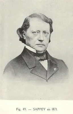
1874 –1875 SAPPEY, Marie Philibert Constant (1810 – 1896)
Anatomie, physiologie, pathologie des vaisseaux lymphatiques.
Notable for its illustrations.
GMN 1110 · https://historyofmedicine.com/id/1179
1881 –1884 RETZIUS, Magnus Gustaf (1842 – 1919)
Das Gehörorgan der Wirbelthiere. 2 vols.
The most magnificent of all comparative anatomical studies of the ear, and the most beautiful studies of the ear after those of Casseri (No.1540). Retzius described the “Retzius bodies” in the labyrinth.
GMN 1566 · https://historyofmedicine.com/id/1237
1884 –1889 BRAUNE, Christian Wilhelm (1831 – 1892)
Das Venensystem des menschlichen Körpers. 2 pts. and atlas.
Like Braune’s other anatomical works, this is notable for its excellent illustrations.
GMN 784 · https://historyofmedicine.com/id/1770
1886 KOLLMANN, Julius Konstantin Ernst (1834 – 1918)
Plastische Anatomie des menschlichen Körpers.
“Illustrated with lithographs from hand-drawings, photographs from the nude, ethnic studies of facial features…The text…is of unusual historic interest, and includes special chapters on the anatomy of the infant, huma…
GMN 427 · https://historyofmedicine.com/id/1023
1889 MONDEVILLE, Henri de (1260 – 1316); PAGEL, Julius Leopold (1851 – 1912)
Die Anatomie des Heinrich von Mondeville. Nach einer Handschrift der Königlichen Bibliothek zu Berlin von Jahre 1304 zum ersten Male herausgegeben von J. Pagel.
Mondeville was the first teacher known to have lectured with the aid of illustrations, using 13 charts of human anatomy. He lectured at Montpellier. Digital facsimile from the Medical Heritage Library, Internet Archiv…
GMN 362 · https://historyofmedicine.com/id/382
1898 –1901 LEONARDO DA VINCI (1452 – 1519); SABACHNIKOFF, Theodore (Fyodor Vasilyevich Sabašnikov) (1869 – 1927); PIUMATI, Giovanni (1850 – 1915)
I manoscritti de Leonardo da Vinci della Reale Biblioteca di Windsor. Pubblicata da Teodoro Sabachnikoff. Transcritti e annotati da Giovanni Piumati. 2 vols.
Includes folios A & B of his anatomical MSS. Text in French and Italian.
GMN 364 · https://historyofmedicine.com/id/416
1908 SUDHOFF, Karl Friedrich Jakob (1853 – 1938)
Ein Beitrag zur Geschichte der Anatomie im Mittelalter, speziell der anatomischen Graphik nach Handschriften des 9. bis 15. Jahrhunderts.
Studien zur Geschichte der Medizin, Leipzig, Heft 4. Reprinted Hildesheim, 1964.
GMN 452 · https://historyofmedicine.com/id/1155
1908 WEINDLER, Fritz
Geschichte der gynäkologische-anatomischen Abbildungen.
From the ancient world through the 18th century. Digital facsimile from Google Books at this link.
GMN 6293 · https://historyofmedicine.com/id/8026
1911 –1916 LEONARDO DA VINCI (1452 – 1519); FONAHN, Adolf Mauritz (1873 – 1940); VANGENSTEN, Ove Conrad Langaard (1880 – 1946); HOPSTOCK, Halfdan (1866 – 1925)
Leonardo da Vinci: Quaderni d’anatomia I-VI. Fogli della Royal Library di Windsor, pubblicati da Ove C.L. Vangensten, A. Fonahn, H. Hopstock. 6 vols.
Leonardo, “the greatest artist and scientist of the Italian Renaissance, was the founder of iconographic and physiologic anatomy” (Garrison). He made over 750 sketches of all the principal organs of the body, drawings…
GMN 365 · https://historyofmedicine.com/id/422
1926 MONDINO DE LUZZI, [MUNDINUS] (1270 – 1326); WICKERSHEIMER, Charles Adolphe Ernest (1880 – 1965); GUIDO DA VIGEVANO, (Guido da Pavia) (1280 – 1349)
Anatomies de Mondino dei Luzzi et de Guido de Vigevano. Par Ernest Wickersheimer.
Facsimile of the 1478 edition of Mondino's Anothomia along with the text and 18 plates from Guido de Vigevano's (fl. 14th century) Anathomia. Vigevano's manuscript, completed in 1345, is MS. 569 in the Musée Condé at …
GMN 8362 · https://historyofmedicine.com/id/10538
1937 –1960 PERNKOPF, Eduard (1888 – 1955)
Topographische Anatomie des Menschen. Lehrbuch und Atlas der regionär-stratigraphischen Präparation. 4 vols. in 7. Vol. 1 in 2 pts: Brust un Brustgliedmasse, 1937; Vol. 2 in 2 pts: Bauch, Becken und Beckengliedmasse, 1941; Vol. 3: Der Hals, 1952; Vol. 4 in 2 pts: Topographische und stratigraphischen Anatomie des Kopfes, 1957, 1960.
Pernkopf's anatomy is remarkable for the intricacy of its detailed images and its "regional stratigraphic" approach, i.e. "multiple layers of dissection with an emphasis on fascia shown and reflected, approaching the …
GMN 10200 · https://historyofmedicine.com/id/12389
1943 VESALIUS, Andreas (1514 – 1564); CUSHING, Harvey Williams (1869 – 1939)
A bio-bibliography of Andreas Vesalius.
The standard annotated bibliography of Vesalius's works, known for its unusual system of numbering entries. Posthumously edited for publication by John F. Fulton and Arturo Castiglioni. Digital facsimile of the 1943 e…
GMN 8096 · https://historyofmedicine.com/id/10272

![PIETRO DA CORTONA, [Pietro BERRETINI] (1596 – 1669)](/media/portraits/743)