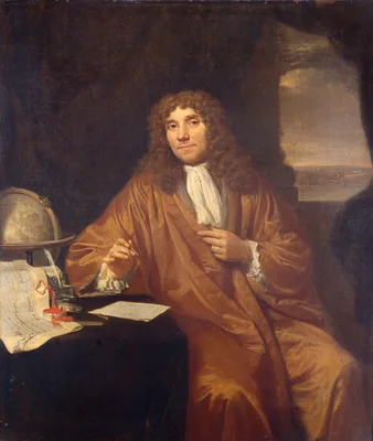
1686 –1718 LEEUWENHOEK, Antonie van (1632 – 1723)
Ontledingen en ontdekkingen.... 6 vols.
Leeuwenhoek, one of the first and also one of the greatest of the microbiologists, communicated many of his discoveries to the Royal Society in London. This set is a collection in Dutch of many contributions that van …
GMN 265 · https://historyofmedicine.com/id/1222
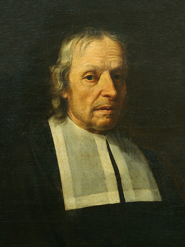
1686 MALPIGHI, Marcello (1628 – 1694)
Opera omnia. 2 vols.
Malpighi was the founder of histology. In 1660 he was the first to see the capillary anastomosis between the arteries and the veins, thus contributing to the completion of Harvey’s work on the circulation. Malpighi wa…
GMN 66 · https://historyofmedicine.com/id/8778
1800 BICHAT, Marie François Xavier (1771 – 1802)
Traité des membranes en général et diverses membranes en particulier.
Bichat conceived the idea of a science of anatomy and pathology based upon an accurate classification of the various tissues of the body, their distribution in the various organs and parts, and their particular suscep…
GMN 537 · https://historyofmedicine.com/id/164
1801 BICHAT, Marie François Xavier (1771 – 1802)
Anatomie générale, appliquée à la physiologie et à la médecine. 4 vols.
Bichat revolutionized descriptive anatomy. Where Morgagni and others had conceived of whole organs being diseased, Bichat showed how individual tissues could be separately affected. He covered tissue pathology, system…
GMN 403 · https://historyofmedicine.com/id/820
1830 MÜLLER, Johannes Peter (1801 – 1858)
De glandularum secernentium structura penitiori.
Müller’s most important histological work. In it he described the microscopic anatomy of a large series of secreting glands. Müller’s greatest influence was not so much through his own work as through the influence he…
GMN 538 · https://historyofmedicine.com/id/166
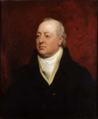
1830 HOME, Sir Everard (1756 – 1832)
A short tract on the formation of tumours, and the peculiarities that are met with in the structure of those that have become cancerous; with their mode of treatment.
Contains the first illustrations of microscopic sections of cancer; however, Home drew no worthwhile conclusion from his microscopic studies. Digital facsimile from the Internet Archive at this link.
GMN 2611 · https://historyofmedicine.com/id/3093
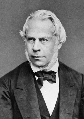
1837 HENLE, Friedrich Gustav Jakob (1809 – 1885)
Symbolae ad anatomiam villorum intestinalium, imprimis eorum epithelii et vasorum lacteorum.
Henle first described the epithelia of the skin and intestines, and defined the structure and function of columnar and ciliated epithelium. He applied the term “epithelium” to all mucous membranes in the body. Modern …
GMN 539 · https://historyofmedicine.com/id/170
1838 –1857 MANDL, Louis (1812 – 1881)
Anatomie microscopique. Tome premier: Histologie. Atlas de cinquante-deux planches. Tome second: Histogénèse. Atlas de quarante planches. 2 vols.
This work was issued in parts over nearly 20 years. Parts concerned specific subjects and were sold separately. Digital facsimile from Google Books at this link, and at this link.
GMN 11117 · https://historyofmedicine.com/id/13313
1841 HENLE, Friedrich Gustav Jakob (1809 – 1885)
Allgemeine Anatomie. Lehre von den Mischungs- und Formbestandtheilen des menschlichen Körpers.
Many of the histological discoveries of Henle are described in the above. He classified tissues histologically. In the section on Gefässnerven (pp. 510, 690) Henle demonstrated the presence of smooth muscle in the end…
GMN 543 · https://historyofmedicine.com/id/268
1845 LEBERT, Hermann (1813 – 1878)
Physiologie pathologique. 2 vols. & atlas.
One of the earliest atlases of pathological histology. Lebert’s work played an important role in introducing the cellular idea of pathology, laying the groundwork for Virchow’s theories.
GMN 543.1 · https://historyofmedicine.com/id/179
1846 –1849 HASSALL, Arthur Hill (1817 – 1894)
The microscopic anatomy of the human body, in health and disease.
First English textbook on microscopical anatomy. His description of the concentric corpuscles of the thymus (p. 9) led to the term “Hassall’s corpuscles”.
GMN 544 · https://historyofmedicine.com/id/180
1850 –1855 QUEKETT, John Thomas
Descriptive and illustrated catalogue of the histological series contained in the Museum of the Royal College of Surgeons of England. Vol. I. Elementary tissues of vegetables and animals; Vol. II. Structure of the skeleton of vertebrate animals. 2 vols.
Digital facsimile from the Hathi Trust at this link.
GMN 13157 · https://historyofmedicine.com/id/15416
1852 KÖLLIKER, Rudolph Albert von (1817 – 1905)
Handbuch der Gewebelehre des Menschen.
Isolation of smooth muscle.
GMN 546 · https://historyofmedicine.com/id/190
1858 GERLACH, Joseph von (1820 – 1896)
Mikroskopische Studien aus dem Gebiete der menschlichen Morphologie.
Gerlach introduced several staining methods, the most important of which (a transparent solution of ammonia carmine and gelatin) is called “Gerlach’s stain”; it was the first satisfactory histological stain.
GMN 548 · https://historyofmedicine.com/id/194
1858 SCHULTZE, Max Johann Sigismund (1825 – 1874)
Ueber die Endigungsweise des Hörnerven im Labyrinth.
Schultze’s great monographs on the nerve-endings of the sense organs were of prime importance in the development of the science of histology. Besides that dealing with the internal ear, he wrote others dealing with th…
GMN 1561 · https://historyofmedicine.com/id/1221
1861 HIS, Wilhelm Sr. (1831 – 1904)
Untersuchungen über den Bau der Lymphdrüsen.
Histology of the lymphatics. His drew the illustrations.
GMN 1107 · https://historyofmedicine.com/id/1166
1862 SCHULTZE, Max Johann Sigismund (1825 – 1874)
Untersuchungen über den Bau der Nasenschleimhaut, namentlich die Structur und Endigungsweise der Geruchsnerven bei dem Menschen und den Wirbelthieren.
Schultze’s classic paper on the nerves to the neuro-epithelium in the special sense organs marks an epoch in histology. He described the cells of the olfactory muccous membrane, “Schultze’s cells”.
GMN 936 · https://historyofmedicine.com/id/175
1863 RECKLINGHAUSEN, Friedrich Daniel von (1833 – 1910)
Ueber Eiter- und Bindegewebskörperchen.
Recklinghausen described granular cells in the frog mesentery, later named “mast cells” by Ehrlich (No. 553.1).
GMN 548.1 · https://historyofmedicine.com/id/200
1864 DEAN, John (1831 – 1888)
The gray substance of the medulla oblongata and trapezium.
The first American medical book illustrated with photomechanically reproduced plates. Oliver Wendell Holmes praised the book for its remarkable photomicrographs, which may be the first published of brain cross-section…
GMN 7331 · https://historyofmedicine.com/id/9502
1869 KLEBS, Theodor Albrecht Edwin (1834 – 1913)
Die Einschmelzungs-Methode, ein Beitrag zur mikroskopischen Technik.
Introduction of paraffin embedding
GMN 549 · https://historyofmedicine.com/id/205
1869 –1872 STRICKER, Salomon (1834 – 1898)
Handbuch der Lehre von den Geweben des Menschen und der Thiere. Edited by Salomon Stricker. 2 vols.
Digital facsimile from the Internet Archive at this link. Translated into English by Henry Power as Manual of human and comparative histology. Edited by S. Stricker. 3 vols. London: New Sydenham Society, 1870-73. Digi…
GMN 550 · https://historyofmedicine.com/id/208
1869 –1876 RANVIER, Louis-Antoine (1835 – 1922); CORNIL, André Victor (1837 – 1908)
Manuel d’histologie pathologique. 3 pts.
English translations, Philadelphia, 1880, and London, 1882-86.
GMN 2300 · https://historyofmedicine.com/id/2937
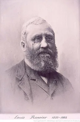
1878 RANVIER, Louis-Antoine (1835 – 1922)
Leçons sur l’histologie du système nerveux. 2 vols.
Includes his description of the “nodes of Ranvier”, interruptions of the medullary nerve sheaths.
GMN 1276 · https://historyofmedicine.com/id/2273
1879 DUVAL, Mathias-Marie (1844 – 1907)
Technique de l’emploi du collodion humide pour la pratique des coupes microscopiques.
Introduction of collodion for embedding.
GMN 553 · https://historyofmedicine.com/id/223
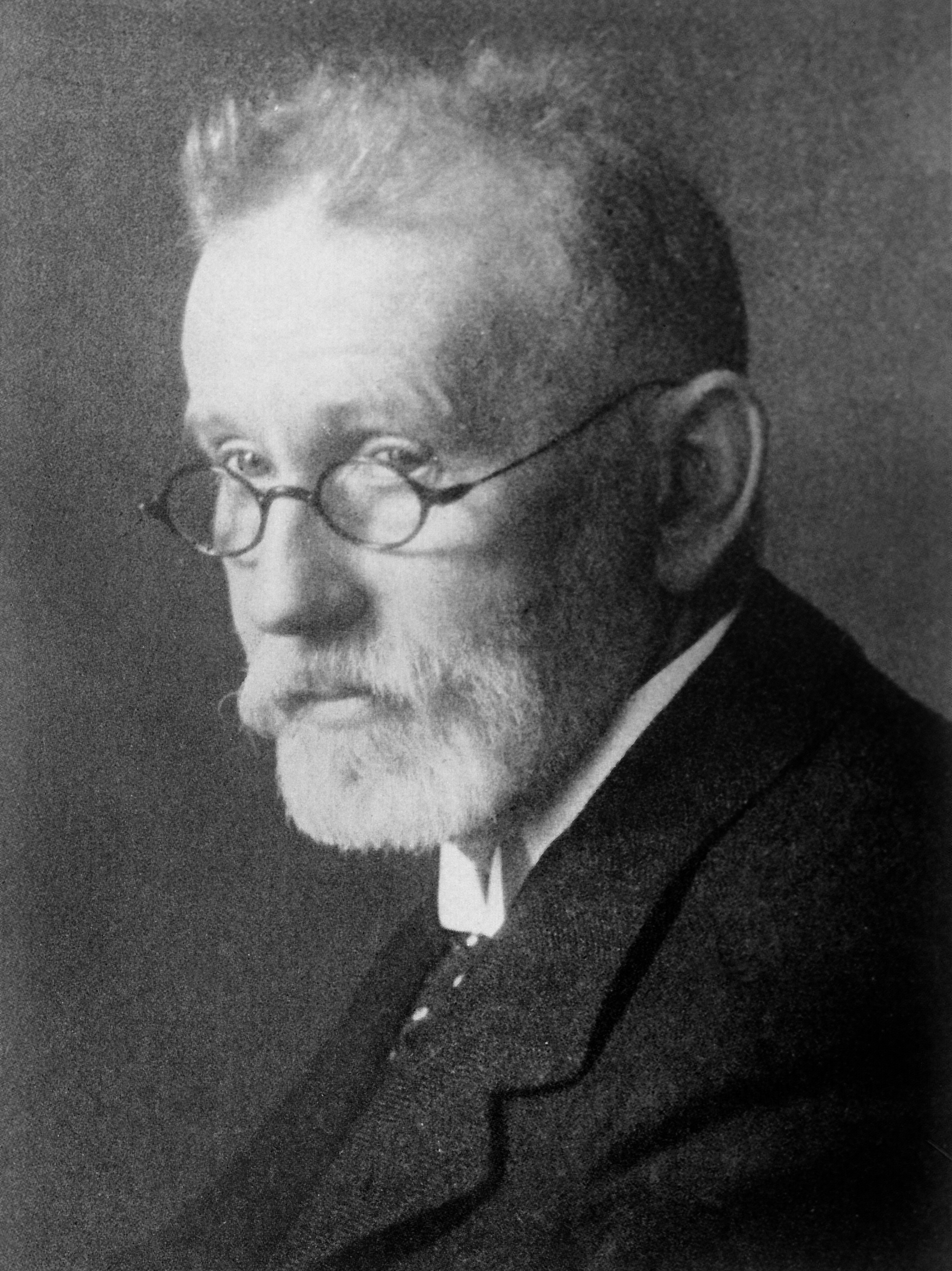
1886 EHRLICH, Paul (1854 – 1915)
Ueber die Methylenblaureaction der lebenden Nervensubstanz.
Ehrlich’s method of intravital staining.
GMN 554 · https://historyofmedicine.com/id/231
1886 GOLGI, Camillo (1844 – 1926)
Sulla fina anatomia degli organi centrali del sistema nervoso.
Golgi’s histological studies made a clear conception of the nervous system possible for the first time. He demonstrated the existence of multipolar nerve-cells (Golgi cells) by means of his silver nitrate stain, and d…
GMN 1416 · https://historyofmedicine.com/id/2103
1892 CAJAL, Santiago Ramón y (1852 – 1934)
Nuevo concepto de la histologia de los centros nerviosos.
Ramón y Cajal, son of a struggling Aragonese doctor, lived to become one of the greatest of all histologists. He devised many staining methods for nervous tissue and did work of fundamental importance to neuroanatomy.…
GMN 1287 · https://historyofmedicine.com/id/2315
1893 BLUM, Ferdinand (1865 – 1859)
Der Formaldehyd als Härtungsmittel. Vorläufige Mittheilung.
Formalin first used for tissue fixation.
GMN 557 · https://historyofmedicine.com/id/261
1905 –1906 TYZZER, Ernest Edward (1875 – 1965)
The histology of the skin lesions in varicella.
Tyzzer was first to recognize inclusion bodies in varicella.
GMN 5440 · https://historyofmedicine.com/id/7464
1913 –1914 CAJAL, Santiago Ramón y (1852 – 1934)
Estudios sobre la degeneración y regeneración de sistema nervioso.
The most complete work on the subject so far written. Ramón y Cajal, great neuroanatomist and histologist, was for many years in charge of the institute bearing his name at Madrid. He gained the Nobel Prize in 1906. E…
GMN 560.1 · https://historyofmedicine.com/id/287
1916 DAWSON, James Walker (1870 – 1927)
The histology of disseminated sclerosis.
A classic monograph on the pathology of multiple sclerosis.
GMN 4718 · https://historyofmedicine.com/id/1201
1916 GASKELL, Walter Holbrook (1847 – 1914)
The involuntary nervous system. Part 1.
This book sums up the life work of Gaskell, who laid the histological foundation of the modern study of the autonomic nervous system. No more published.
GMN 1331 · https://historyofmedicine.com/id/1424
1919 MUMMERY, John Howard (1847 – 1926)
The microscopic anatomy of the teeth.
Digital facsimile of the 1919 edition from Biodiversity Heritage Library at this link. Mummery substantially expanded and retitled the second edition of this work as The microscopic & general anatomy of the teeth huma…
GMN 12507 · https://historyofmedicine.com/id/14741
1920 MACEWEN, Sir William (1848 – 1924)
The growth and shedding of the antlers of the deer. The histological phenomena and their relation to the growth of bone.
The first study of the unusual and dramatic physiology of the annual growth and shedding of the antlers of deer. "The amount of bony matter annually secreted to form antlers of the larger deer is enormous, antlers of …
GMN 7000 · https://historyofmedicine.com/id/9166
1926 –1978 HENKE, Friedrich (1868 – 1943); LUBARSCH, Otto (1860 – 1933); RÖSSLE, Robert (1876 – 1856)
Handbuch der speziellen pathologischen Anatomie und Histologie. Edited by Friedrich Henke, Otto Lubarsch and Robert Rössle. 13 vols. in 43.
1: Blut, Knochenmark, Lymphknoten, Milz. 1/1: Blut, Lymphknoten von Max Askanazy. 1926. 1/2: Milz, Knochenmark von Max Askanazy. 1927. 3A: Lymphknoten: Diagnostik in Schnitt und Ausstrich: Cytologie und Lymphadenitis …
GMN 2311 · https://historyofmedicine.com/id/2967
1941 POLYAK, Stephen Lucian (1889 – 1955)
The anatomy and the histology of the retina in man, ape, and monkey.
A scholarly tour-de-force with a bibliography of over 700 references.
GMN 1533.1 · https://historyofmedicine.com/id/2860
1956 –1960 EHRLICH, Paul (1854 – 1915); HIMMELWEIT, Fred (1902 – 1977)
Collected papers of Paul Ehrlich. Compiled and edited by F. Himmelweit. 3 vols.
Vol. I: Histology, biochemistry, and pathology, Vol. 2: Immunology and cancer research; Vol. 3: Chemotherapy. Most texts are in German. English translations are also published when available. The set includes new Engl…
GMN 86.4 · https://historyofmedicine.com/id/1453
1971 HOGAN, Michael J. (1907 – 1976); ALVARADO, Jorge A.; WEDDELL, Joan Esperson
Histology of the human eye.
Hogan and Alvarado's work was the first book on histology of the eye to include electron microscopy. It also reproduced spectacular three-dimensional representations of ocular ultrastructures by Joan Esperson Weddell.
GMN 7405 · https://historyofmedicine.com/id/9577