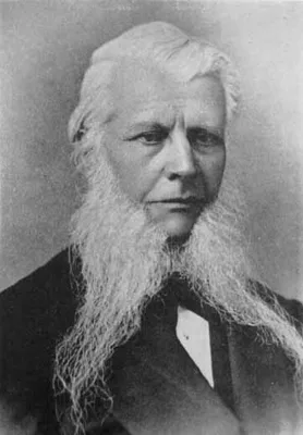
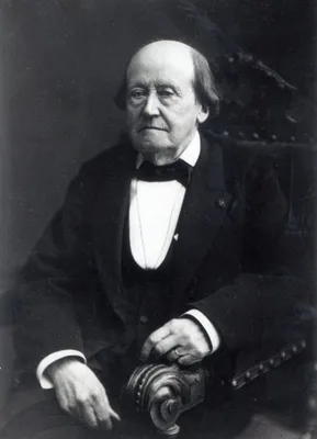
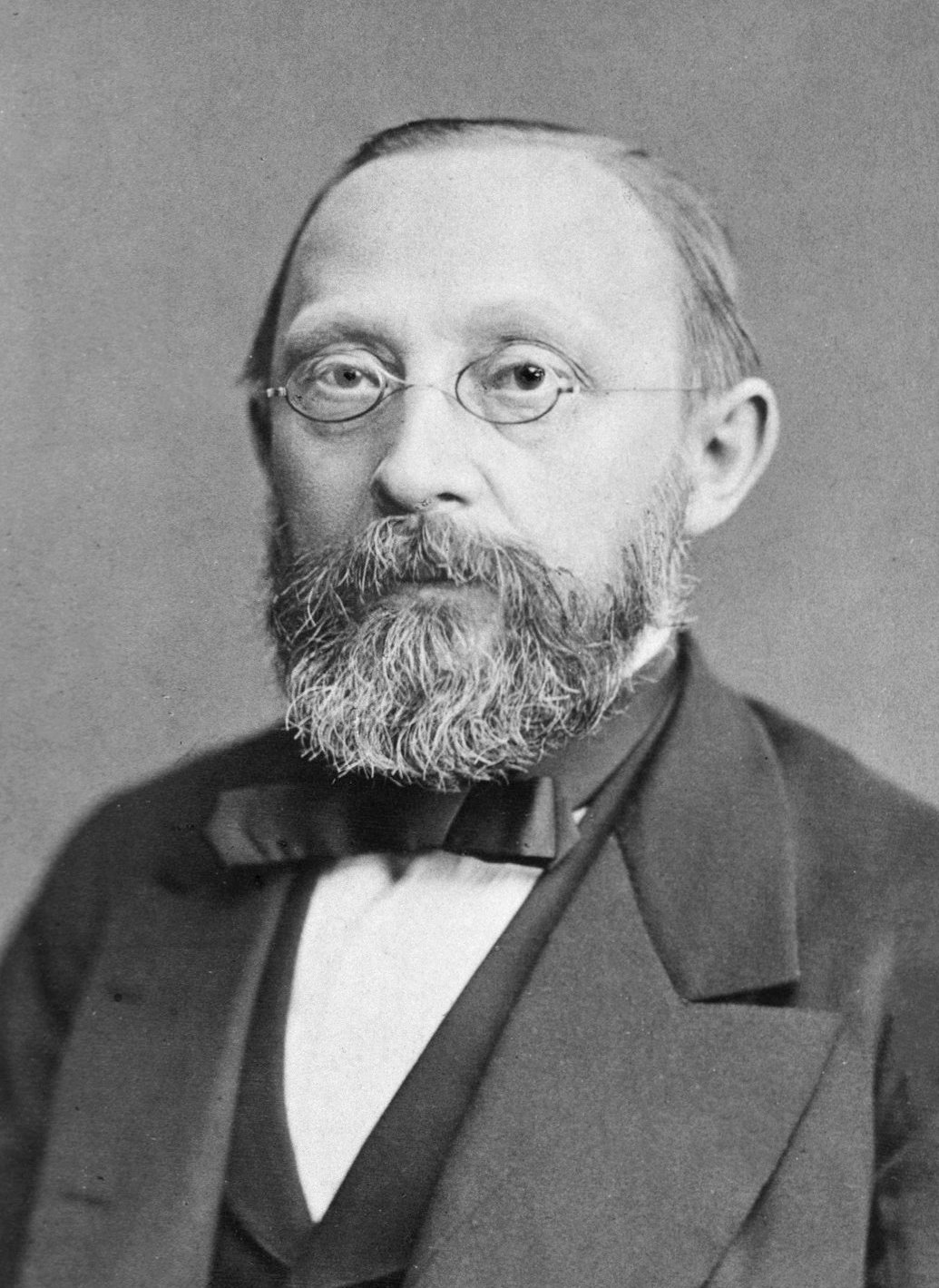
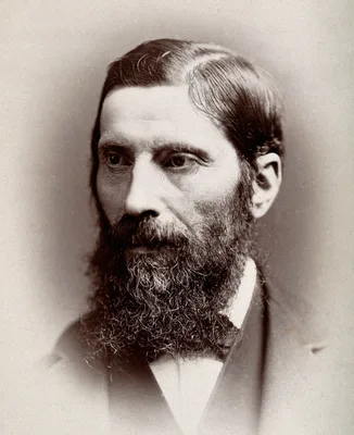
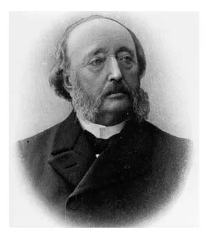
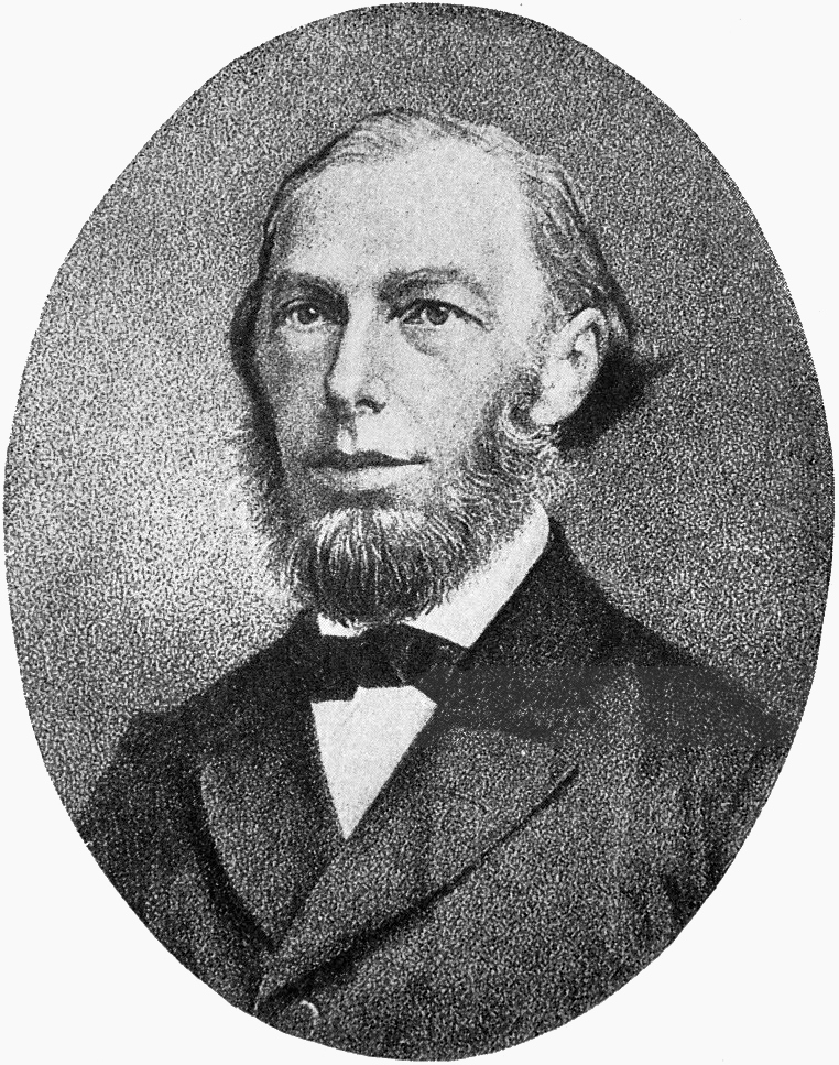
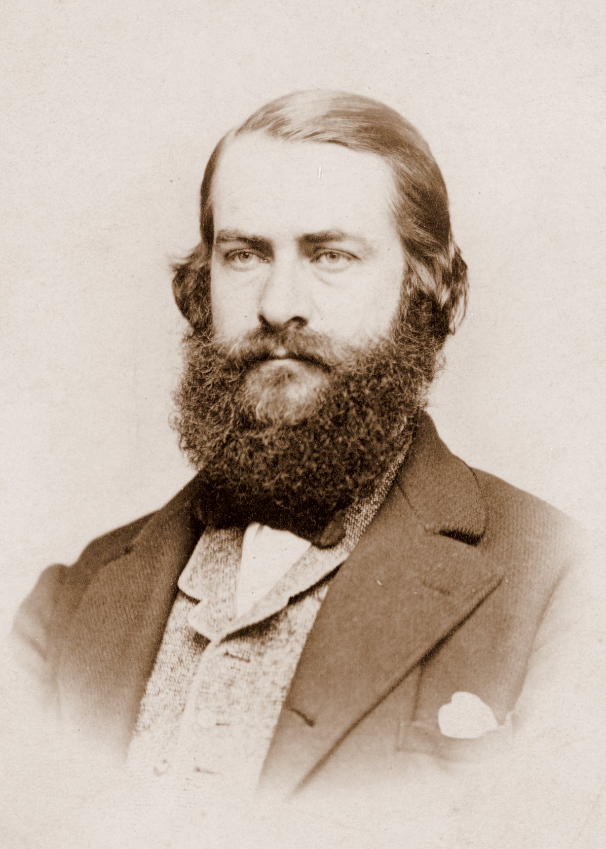
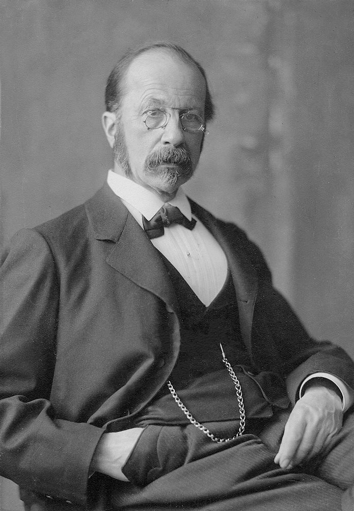
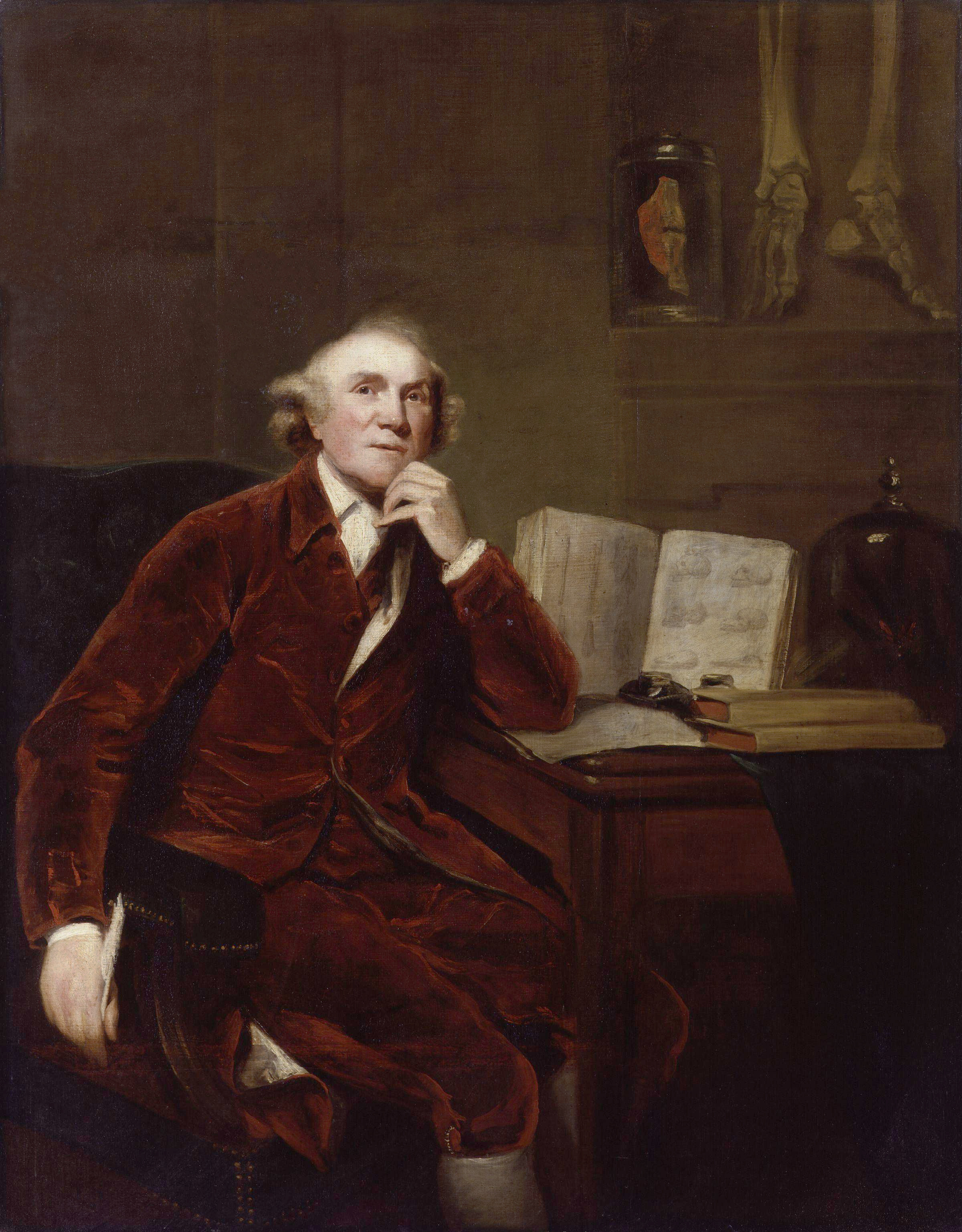
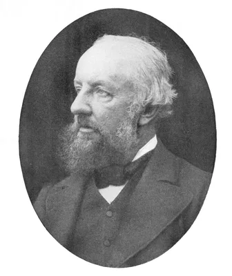
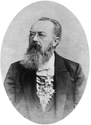
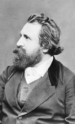
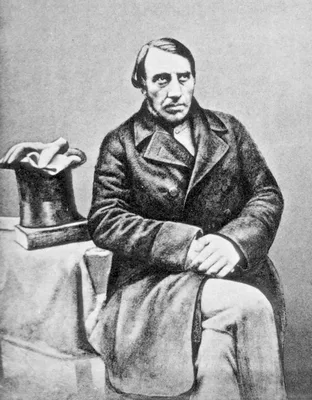
1852 BIDDER, Friedrich Karl Heinrich von (1810 – 1894)
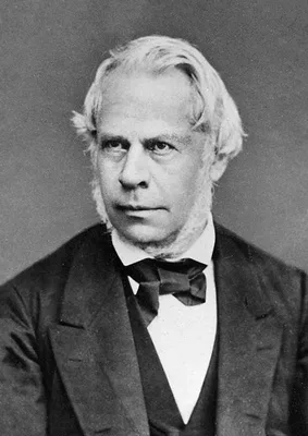
Ueber functionell verschiedene und räumlich getrennte Nervencentra im Froschherzen.
Discovery of the ganglion cells at the auriculo-ventricular junction, “Bidder’s ganglion”.
GMN 811 · https://historyofmedicine.com/id/203
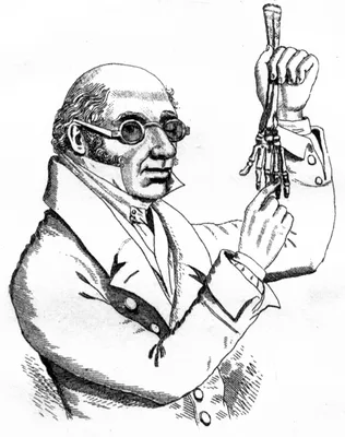
1852 KNOX, Robert (1791 – 1862)
A manual of artistic anatomy.
Knox, remembered because of his indiscreet association with the Edinburgh “resurrectionists”, was one of the best teachers of anatomy during the 19th century.
GMN 415 · https://historyofmedicine.com/id/945
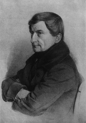
1852 –1859 PIROGOV, Nikolai Ivanovich (Pirogoff) (1810 – 1881)
Anatome topographica sectionibus per corpus humanum congelatum triplici directione ductis illustrata. 8 pts.
Pirogov was the greatest of Russian surgeons. He introduced the teaching of applied topographical anatomy in Russia. His atlas of 220 plates represents the first use on a grand scale of frozen sections in anatomical i…
GMN 416 · https://historyofmedicine.com/id/950
1852 CHOULANT, Johann Ludwig (1791 – 1861)
Geschichte und Bibliographie der anatomischen Abbildung.
In this classic work Choulant traced the evolution of anatomical illustration from the early schematic plates up to his own time, including a valuable bibliography. Reprinted, Wiesbaden, 1974. An English translation b…
GMN 440 · https://historyofmedicine.com/id/1098
1853 TREITZ, Václav (Wenzel) (1819 – 1872)
Ueber einen neuen Muskel am Duodenum des Menschens, über elastische Sehnnen, und einige andere anatomische Verhãltnisse.
Treitz, a Czech pathologist, discovered the muscle at the duodenojejunal junction, later called “muscle of Treitz”; the fold of peritoneum over the muscle of Treitz is known as the "ligament of Treitz."
GMN 7241 · https://historyofmedicine.com/id/9410
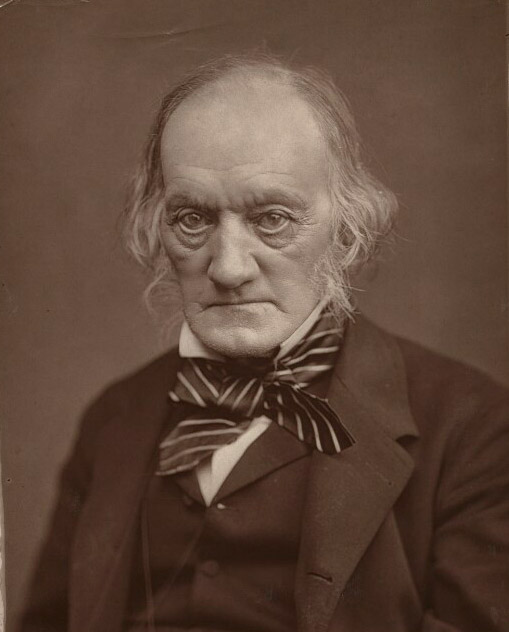
1853 OWEN, Sir Richard (1804 – 1892)
Descriptive catalogue of the osteological series contained in the museum of the Royal College of Surgeons of England. 2 vols.
Digital facsimile from wellcomecollection.org at this link.
GMN 13860 · https://historyofmedicine.com/id/16150
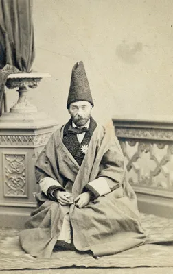
1854 MIRZA MOHAMMAD-VALI HAKIMBASHI
[In Persian script] Cheragh haa rewshenaaa der asewl pezeshekea [Illumination of the fundamentals of medicine].
Issued in 1271 A. H. (1854 CE), this entirely lithographed book introduced Western anatomical illustration to Persian culture. As part of an effort to modernize medical education in Persia, medical textbooks such as M…
GMN 6833 · https://historyofmedicine.com/id/8997
1854 POLAK, Jakob Eduard (1818 – 1891)
Kitab fi tashrih beden al-insan [in Persian; English translation: Anatomy of the human body]. Lithographed text.
The first original Persian-language anatomy textbook based on western medical science, printed in a very small number of copies for the use of Polak’s Persian students. Polak, an Austrian physician, was responsible fo…
GMN 14061 · https://historyofmedicine.com/id/16371
1855 –1871 HENLE, Friedrich Gustav Jakob (1809 – 1885)
Handbuch der systematischen Anatomie des Menschen. 3 vols.
Considered by many authorities to be the greatest of the 19th-century systems of anatomy. Many structures are named after Henle, including the looped portion of the uriniferous tubules of the kidney, the layer of cell…
GMN 417 · https://historyofmedicine.com/id/953
1855 –1869 SIBSON, Francis (1814 – 1876)
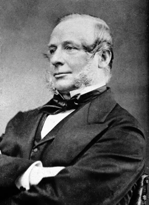
Medical anatomy: or, illustrations of the relative position and movements of the internal organs. 7 pts.
Sibson was professor of medicine at St. Mary’s Hospital. “Sibson’s fascia” and “muscle” are named after him. Plates 19-21 show movements, structure and sounds of the heart.
GMN 422 · https://historyofmedicine.com/id/999
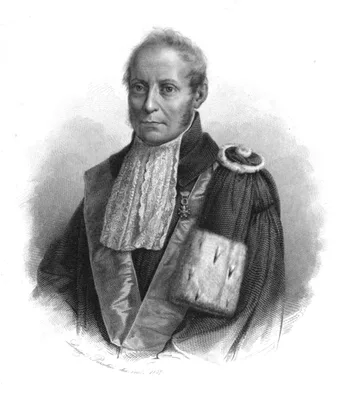
1857 MEDICI, Michele (1782 – 1859)
Compendio storico della scuola anatomica di Bologna dal Rinascimento delle scienze e delle letters a tutto il secolo XVIII. Con un paragone fra la sua antichità e quella delle scuole di Salerno e di Padova.
Digital facsimile from Google Books at this link.
GMN 8854 · https://historyofmedicine.com/id/11032
1857 –1881 MILNE-EDWARDS, Henri (1800 – 1885)
Leçons sur la physiologie et l'anatomie comparée de l'homme et des animaux faites a la Faculté des Sciences de Paris. 14 vols.
Digital facsimile of all the volumes from Google Books; vol. 1 at this link.
GMN 12016 · https://historyofmedicine.com/id/14224
1857 VIRCHOW, Rudolf Ludwig Karl (1821 – 1902)
Untersuchungen uber die Entwickelung des Schädelgrundes im gesunden und krankhaften Zustande und über Einfluss derselben auf Schädelform, Gesichtsbildung und Gehirnbau.
In his Investigations on the development of the base of the skull in healthy and diseased conditions, and on the influence of the same upon skull form, facial structure and brain formation Virchow laid the foundation …
GMN 13248 · https://historyofmedicine.com/id/15513
1858 GERLACH, Joseph von (1820 – 1896)
Mikroskopische Studien aus dem Gebiete der menschlichen Morphologie.
Gerlach introduced several staining methods, the most important of which (a transparent solution of ammonia carmine and gelatin) is called “Gerlach’s stain”; it was the first satisfactory histological stain.
GMN 548 · https://historyofmedicine.com/id/194
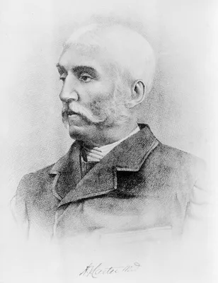
1858 GRAY, Henry (1825 – 1861); CARTER, Henry Vandyke (1831 – 1897)
Anatomy, descriptive and surgical
Gray’s textbook of anatomy remains today a standard work on the subject in the English-speaking world. The 37th edition appeared in 1989; the first American edition was published at Philadelphia, 1859. Digital facsimi…
GMN 418 · https://historyofmedicine.com/id/957
1858 HUMPHRY, Sir George Murray (1820 – 1896)
A treatise on the human skeleton, including the joints.
Humphry was professor of anatomy at Cambridge and became the first professor of surgery there. He founded the Journal of Anatomy and Physiology in 1867. “Humphry’s ligament” of the knee-joint is described on p. 546 of…
GMN 419 · https://historyofmedicine.com/id/959
1858 OLLIER, Louis Xavier Édouard Léopold (1830 – 1900)
Des moyens chirurgicaux de favoriser la reproduction des os après les résections.
“Ollier’s layer”, the osteogenetic layer of the periosteum.
GMN 420 · https://historyofmedicine.com/id/993
1858 SCHULTZE, Max Johann Sigismund (1825 – 1874)
Ueber die Endigungsweise des Hörnerven im Labyrinth.
Schultze’s great monographs on the nerve-endings of the sense organs were of prime importance in the development of the science of histology. Besides that dealing with the internal ear, he wrote others dealing with th…
GMN 1561 · https://historyofmedicine.com/id/1221
1858 LE GENDRE, Eugène-Quintien (1823 – )
Anatomie chirurgicale homolographique, ou Déscription et figures des principales regions du corps humain, representées de grandeur naturelle, d'après des sections planes pratiquées sur des cadavres congeles....
Contains 25 plates lithographed by Lemercier after drawings by the author representing natural size sections taken in horizontal, sagittal and oblique planes from various portions of the body. The first plate of the s…
GMN 13193 · https://historyofmedicine.com/id/15454
1859 STILLING (BENEDICT), Benedikt (1810 – 1879)
Neue Untersuchungen über den Bau des Rückenmarks. 2 vols. (Text and atlas.)
Stilling carried out some of the 19th century’s most detailed and precise examinations of the spinal cord, which “laid the foundation for the modern anatomical study of the spinal cord, medulla oblongata, and pons” (C…
GMN 14214 · https://historyofmedicine.com/id/16530
1861 LEIDY, Joseph (1823 – 1891)
An elementary treatise on human anatomy.
Leidy illustrated this book himself. He was professor of anatomy at Philadelphia and the leading American anatomist of his time.
GMN 421 · https://historyofmedicine.com/id/997
1861 HIS, Wilhelm Sr. (1831 – 1904)
Untersuchungen über den Bau der Lymphdrüsen.
Histology of the lymphatics. His drew the illustrations.
GMN 1107 · https://historyofmedicine.com/id/1166
1861 HUNTER, John (1728 – 1793); OWEN, Sir Richard (1804 – 1892)
Essays and observations on natural history, anatomy, physiology, psychology, and geology by John Hunter, F.R.S. Being his posthumous papers on those subjects, arranged and revised, with notes; to which are added the introductory lectures on the Hunterian collection of fossil remains delivered in the theatre of the Royal College of Surgeons of England, March 8th, 10th and 12th, 1855 by Richard Owen .... 2 vols.
Digital facsimiles from the Hathi Trust at this link.
GMN 7608 · https://historyofmedicine.com/id/9780
1861 –1867 RÜDINGER, Nikolaus (1832 – 1896)
Atlas des peripherischen Nervensystems des menschlichen Körpers / Atlas du système nerveux périphérique du corps humain. Mit einem Vorwort von Prof. Dr. Th. W. L. Bischoff, nach der Natur photographirt von Joseph Alb. 10 parts.
Issued in 10 parts in folio (510 x 360 mm). Includes 46 large-format original photographs mounted on sheets of cardboard.
GMN 14159 · https://historyofmedicine.com/id/16474
1862 SCHULTZE, Max Johann Sigismund (1825 – 1874)
Untersuchungen über den Bau der Nasenschleimhaut, namentlich die Structur und Endigungsweise der Geruchsnerven bei dem Menschen und den Wirbelthieren.
Schultze’s classic paper on the nerves to the neuro-epithelium in the special sense organs marks an epoch in histology. He described the cells of the olfactory muccous membrane, “Schultze’s cells”.
GMN 936 · https://historyofmedicine.com/id/175
1862 RECKLINGHAUSEN, Friedrich Daniel von (1833 – 1910)
Die Lymphgefässe und ihre Beziehung zum Bindegewebe.
“Recklinghausen’s canals”, the lymph canaliculi.
GMN 1108 · https://historyofmedicine.com/id/1169
1862 CLARK, William (1788 – 1869); CLARK, John Willis (1833 – 1910)
Catalogue of the osteological portion of specimens contained in the Anatomical Museum of the University of Cambridge.
Digital facsimile from the Internet Archive at this link.
GMN 7621 · https://historyofmedicine.com/id/9793
1863 RECKLINGHAUSEN, Friedrich Daniel von (1833 – 1910)
Ueber Eiter- und Bindegewebskörperchen.
Recklinghausen described granular cells in the frog mesentery, later named “mast cells” by Ehrlich (No. 553.1).
GMN 548.1 · https://historyofmedicine.com/id/200
1864 DEAN, John (1831 – 1888)
The gray substance of the medulla oblongata and trapezium.
The first American medical book illustrated with photomechanically reproduced plates. Oliver Wendell Holmes praised the book for its remarkable photomicrographs, which may be the first published of brain cross-section…
GMN 7331 · https://historyofmedicine.com/id/9502
1865 SCHULTZE, Bernhard Sigmund (1827 – 1919)
Wandtafeln zur Schwangerschafts- und Geburtskunde. Text volume in quarto format plus atlas in double elephant folio format (915 x 650mm.)
This huge atlas of obstetric wall charts contains 20 chromolithographed plates measuring over 3 feet by 2 feet, illustrating the female reproductive anatomy, stages of pregnancy, normal and breech presentations of the…
GMN 10273 · https://historyofmedicine.com/id/12464
1867 MEYNERT, Theodor Hermann (1833 – 1892)
Der Bau der Gross-Hirnrinde und seine örtlichen Verschiedenheiten, nebst einem pathologisch-anatomischen Corollarium.
Meynert noticed regional variations in the histological structure of different parts of the gray matter in the cerebral hemispheres. He is credited with beginning the study of cytoarchitecture. Meynert described the f…
GMN 1403 · https://historyofmedicine.com/id/2035
1868 GOODSIR, John (1814 – 1867); TURNER, Sir William (1832 – 1916); LONSDALE, Henry (1816 – 1876)
The anatomical memoirs of John Goodsir F.R.S. Edited by William Turner. With a biographical memoir by Henry Lonsdale. 2 vols.
GMN 13221 · https://historyofmedicine.com/id/15485
1869 KLEBS, Theodor Albrecht Edwin (1834 – 1913)
Die Einschmelzungs-Methode, ein Beitrag zur mikroskopischen Technik.
Introduction of paraffin embedding
GMN 549 · https://historyofmedicine.com/id/205
1869 –1872 STRICKER, Salomon (1834 – 1898)
Handbuch der Lehre von den Geweben des Menschen und der Thiere. Edited by Salomon Stricker. 2 vols.
Digital facsimile from the Internet Archive at this link. Translated into English by Henry Power as Manual of human and comparative histology. Edited by S. Stricker. 3 vols. London: New Sydenham Society, 1870-73. Digi…
GMN 550 · https://historyofmedicine.com/id/208
1869 LANGERHANS, Paul (1847 – 1888)
Beiträge zur mikroskopischen Anatomie der Bauchspeicheldrüse. Inaugural-Dissertation.
First account of the islets of Langerhans. In 1893 Édouard Laguesse attached the name of Langerhans to the structures. Langerhans did not suggest any function for them. The book was reprinted with an English translati…
GMN 1009 · https://historyofmedicine.com/id/821
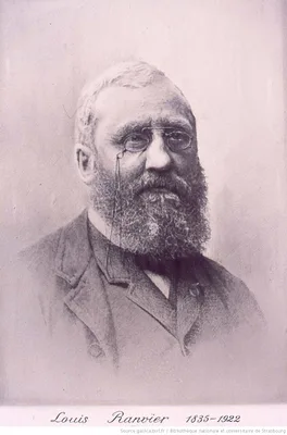
1869 –1876 RANVIER, Louis-Antoine (1835 – 1922); CORNIL, André Victor (1837 – 1908)
Manuel d’histologie pathologique. 3 pts.
English translations, Philadelphia, 1880, and London, 1882-86.
GMN 2300 · https://historyofmedicine.com/id/2937
1870 HEITZMANN, Carl (1836 – 1896)
Die descriptive und topographische Anatomie des Menschen.
GMN 423 · https://historyofmedicine.com/id/1003
1870 KEEN, William Williams (1837 – 1932)
A sketch of the early history of practical anatomy. The introductory address to the course of lectures on anatomy at the Philadelphia School of Anatomy.
Reprinted in 1874 by Lippincott as a separate pamphlet, and in Keen’s Addresses and other papers, Philadelphia, 1905.
GMN 442 · https://historyofmedicine.com/id/1103
1870 LANGHANS, Theodor (1839 – 1915)
Zur Kenntnis der menschlichen Placenta.
“Langhans’s layer”—the cytotrophoblast, the individual cells of which are termed “Langhans’s cells”.
GMN 491 · https://historyofmedicine.com/id/1419
1872 BRAUNE, Christian Wilhelm (1831 – 1892)
Topographisch-anatomischer Atlas. Nach Durchschnitten angefrornen Cadavern.
Fine illustrations of frozen sections. Translated into English by Edward Bellamy as An atlas of topographical anatomy after plane sections of frozen bodies. (Philadelphia: Lindsay and Blakiston, 1877).
GMN 424 · https://historyofmedicine.com/id/1010
1872 BRAUNE, Christian Wilhelm (1831 – 1892)
Die Lage des Uterus und Foetus am Ende der Schwangerschaft nach Durchschnitten an gefrornen Cadavern
Supplement to No. 424.
GMN 6188 · https://historyofmedicine.com/id/7527
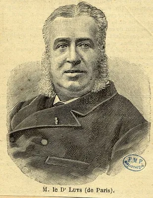
1873 LUYS, Jules Bernard (1828 – 1897)
Iconographie photographique des centres nerveux.
Contains 70 photographs of brain sections taken by Luys himself, with 64 lithographed schemas based on his drawings. Luys undertook this work when the evidence of his lithographs published in 1865 (No. 4012) was dispu…
GMN 1406.01 · https://historyofmedicine.com/id/2066
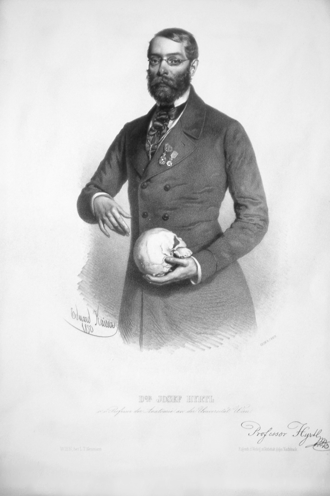
1873 HYRTL, Josef (1810 – 1894)
Die Corrosions-Anatomie und ihre Ergebnisse: mit 18 chromolithographirten Tafeln.
Hyrtl significantly enhanced the techniques of corrosion anatomy, a technique of preparing anatomical specimens invented by Frederik Ruysch. He built up a collection unsurpassed in Europe. In this work Hyrtl described…
GMN 425 · https://historyofmedicine.com/id/9802
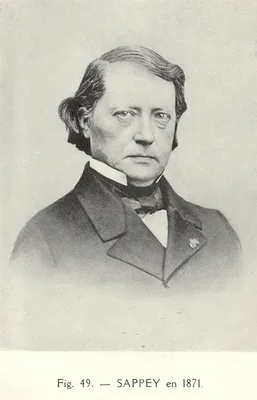
1874 –1875 SAPPEY, Marie Philibert Constant (1810 – 1896)
Anatomie, physiologie, pathologie des vaisseaux lymphatiques.
Notable for its illustrations.
GMN 1110 · https://historyofmedicine.com/id/1179
1875 –1876 RETZIUS, Magnus Gustaf (1842 – 1919); KEY, Axel (1832 – 1901)
Studien in der Anatomie des Nervensystems und des Bindegewebes. Erste Hälfte und zweite Hälfte, erste und zweite Abtheilung. 2 vols.
One of the most strikingly beautiful neuroanatomies ever published, with exquisite reproductions of the color dye injection experiments. The authors confirmed the existence of the foramina of Magendie and Luschka, and…
GMN 1408.2 · https://historyofmedicine.com/id/2079
1876 FISCHER, Ernst
Ueber den Bau der Meissner’schen Tastkörperchen.
Includes first account of the demonstration of nerve endings by means of the gold chloride method.
GMN 551 · https://historyofmedicine.com/id/212
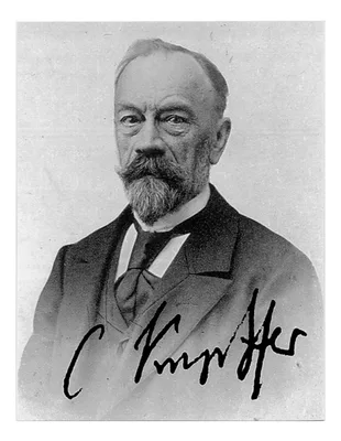
1876 KUPFFER, Karl Wilhelm von (1829 – 1902)
Ueber Stemzellen der Leber.
“Kupffer cells” – macrophage stellate cells in the lining of the blood channels in the liver. Confocal microscopy picture showing the steady-state location and interactions between Kupffer cells (Red), hepatic stellat…
GMN 552 · https://historyofmedicine.com/id/220
1876 VIRCHOW, Rudolf Ludwig Karl (1821 – 1902)
Die Sections-Technik im Leichenhause des Charité-Krankenhauses.
On the technique of dissection. English translation, London, 1876.
GMN 2304 · https://historyofmedicine.com/id/2946
1877 RIMMER, William (1816 – 1879)
Art anatomy.
Perhaps the first great American anatomy for artists by an American painter and sculptor. Rimmer not only drew the 900 drawings on the 81 heliotype plates, but he also wrote in the explanatory text on the sheets along…
GMN 11096 · https://historyofmedicine.com/id/13292
1878 HEIDENHAIN, Rudolf Peter Heinrich (1834 – 1897)
Ueber die Pepsinbildung in den Pylorusdrusen.
Heidenhain pouch.
GMN 1012.1 · https://historyofmedicine.com/id/846