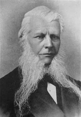
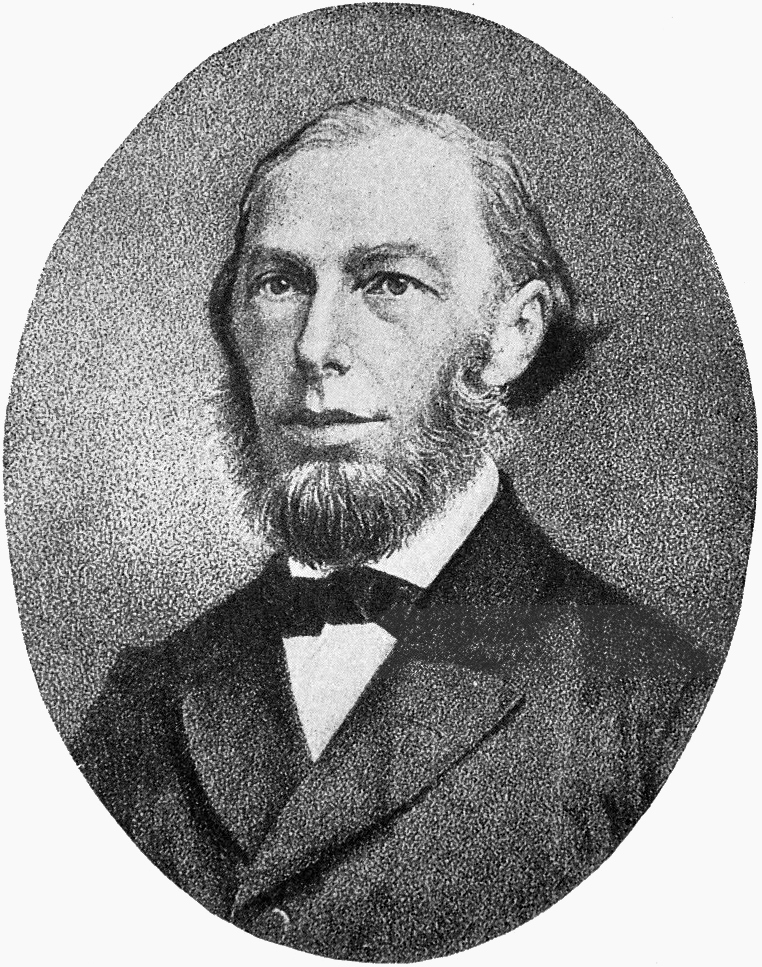
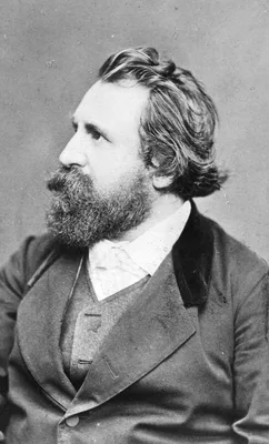
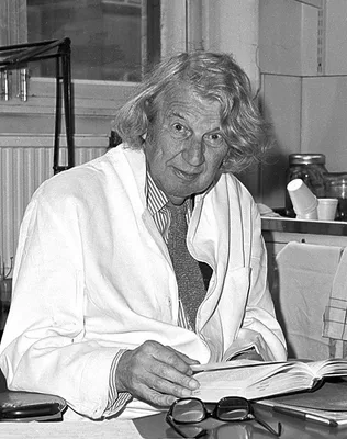
1852 BIDDER, Friedrich Karl Heinrich von (1810 – 1894)
Ueber functionell verschiedene und räumlich getrennte Nervencentra im Froschherzen.
Discovery of the ganglion cells at the auriculo-ventricular junction, “Bidder’s ganglion”.
GMN 811 · https://historyofmedicine.com/id/203
1859 STILLING (BENEDICT), Benedikt (1810 – 1879)
Neue Untersuchungen über den Bau des Rückenmarks. 2 vols. (Text and atlas.)
Stilling carried out some of the 19th century’s most detailed and precise examinations of the spinal cord, which “laid the foundation for the modern anatomical study of the spinal cord, medulla oblongata, and pons” (C…
GMN 14214 · https://historyofmedicine.com/id/16530
1861 –1867 RÜDINGER, Nikolaus (1832 – 1896)
Atlas des peripherischen Nervensystems des menschlichen Körpers / Atlas du système nerveux périphérique du corps humain. Mit einem Vorwort von Prof. Dr. Th. W. L. Bischoff, nach der Natur photographirt von Joseph Alb. 10 parts.
Issued in 10 parts in folio (510 x 360 mm). Includes 46 large-format original photographs mounted on sheets of cardboard.
GMN 14159 · https://historyofmedicine.com/id/16474
1862 SCHULTZE, Max Johann Sigismund (1825 – 1874)
Untersuchungen über den Bau der Nasenschleimhaut, namentlich die Structur und Endigungsweise der Geruchsnerven bei dem Menschen und den Wirbelthieren.
Schultze’s classic paper on the nerves to the neuro-epithelium in the special sense organs marks an epoch in histology. He described the cells of the olfactory muccous membrane, “Schultze’s cells”.
GMN 936 · https://historyofmedicine.com/id/175
1864 DEAN, John (1831 – 1888)
The gray substance of the medulla oblongata and trapezium.
The first American medical book illustrated with photomechanically reproduced plates. Oliver Wendell Holmes praised the book for its remarkable photomicrographs, which may be the first published of brain cross-section…
GMN 7331 · https://historyofmedicine.com/id/9502
1867 MEYNERT, Theodor Hermann (1833 – 1892)
Der Bau der Gross-Hirnrinde und seine örtlichen Verschiedenheiten, nebst einem pathologisch-anatomischen Corollarium.
Meynert noticed regional variations in the histological structure of different parts of the gray matter in the cerebral hemispheres. He is credited with beginning the study of cytoarchitecture. Meynert described the f…
GMN 1403 · https://historyofmedicine.com/id/2035
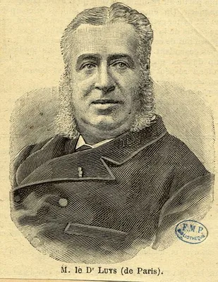
1873 LUYS, Jules Bernard (1828 – 1897)
Iconographie photographique des centres nerveux.
Contains 70 photographs of brain sections taken by Luys himself, with 64 lithographed schemas based on his drawings. Luys undertook this work when the evidence of his lithographs published in 1865 (No. 4012) was dispu…
GMN 1406.01 · https://historyofmedicine.com/id/2066
1875 –1876 RETZIUS, Magnus Gustaf (1842 – 1919); KEY, Axel (1832 – 1901)
Studien in der Anatomie des Nervensystems und des Bindegewebes. Erste Hälfte und zweite Hälfte, erste und zweite Abtheilung. 2 vols.
One of the most strikingly beautiful neuroanatomies ever published, with exquisite reproductions of the color dye injection experiments. The authors confirmed the existence of the foramina of Magendie and Luschka, and…
GMN 1408.2 · https://historyofmedicine.com/id/2079
1876 FISCHER, Ernst
Ueber den Bau der Meissner’schen Tastkörperchen.
Includes first account of the demonstration of nerve endings by means of the gold chloride method.
GMN 551 · https://historyofmedicine.com/id/212
1880 GOLGI, Camillo (1844 – 1926)
Sulla struttura delle fibre nervosa midollate periferiche e centrali.
“Golgi cells” first described.
GMN 1277 · https://historyofmedicine.com/id/2276
1884 MEYNERT, Theodor Hermann (1833 – 1892)
Psychiatrie. Klinik der Erkrankungen der Vorderhirns.
Meynert, Professor of Neurology at Vienna, made many contributions to the study of the cellular architecture of the brain, and is often considered the founder of cerebral cortex cytoarchitectonics. English translation…
GMN 4942 · https://historyofmedicine.com/id/6221
1885 DALTON, John Call (1825 – 1889)
Topographical anatomy of the brain. 3 vols.
The most outstanding American neurological atlas of the nineteenth century and one of the best American photographically illustrated medical books of the period. The atlas reproduces the specimens, which Dalton prepar…
GMN 7332 · https://historyofmedicine.com/id/9503
1887 HIS, Wilhelm Sr. (1831 – 1904)
Zur Geschichte der menschlichen Rückenmarkes und der Nervenwurzeln.
This is generally considered the first clear statement that nerve cells in the brain might be independent units rather than forming an anastomotic network—as Gerlach, Golgi, and most others believed—although there are…
GMN 1368.1 · https://historyofmedicine.com/id/1720
1887 FOREL, Auguste Henri (1848 – 1931)
Einige hirnanatomische Betrachtungen und Ergebnisse.
Independently of His, Forel formulated the neuron theory.
GMN 1368.2 · https://historyofmedicine.com/id/1722
1888 BROCA, Pierre Paul (1824 – 1880)
Mémoires sur le cerveau de l’homme et des primates publiés avec un introduction et des notes par Le docteur S. Pozzi.
Digital facsimile from the Biodiversity Heritage Library at this link.
GMN 344 · https://historyofmedicine.com/id/249
1888 CAJAL, Santiago Ramón y (1852 – 1934)
Estructura de los centros nerviosos de las aves.
"The foundational article of modern cellular neuroscience. . . . this is where Cajal first demonstrated with the Golgi method how neurons interact by contact not continuity in the adult central nervous system—in this …
GMN 7334 · https://historyofmedicine.com/id/9505
1889 HIS, Wilhelm Sr. (1831 – 1904)
Die Neuroblasten und deren Entstehung im embryonalen Mark.
In this paper on neuroblasts (young neurons) and their development in the embryonic spinal cord, His coined the term, “dendrite,” for what had been called protoplasmic processes since the term was introduced by Deiter…
GMN 7335 · https://historyofmedicine.com/id/9506
1891 WALDEYER-HARTZ, Heinrich Wilhelm Gottfried von (1836 – 1921)
Ueber einige neuere Forschungen im Gebiete der Anatomie des Centralnervensystems.
A statement of the neuron theory, to which Waldeyer gave the name.
GMN 1369 · https://historyofmedicine.com/id/1725
1892 CAJAL, Santiago Ramón y (1852 – 1934)
Nuevo concepto de la histologia de los centros nerviosos.
Ramón y Cajal, son of a struggling Aragonese doctor, lived to become one of the greatest of all histologists. He devised many staining methods for nervous tissue and did work of fundamental importance to neuroanatomy.…
GMN 1287 · https://historyofmedicine.com/id/2315
1893 MACEWEN, Sir William (1848 – 1924)
Atlas of head sections. Fifty-three engraved copperplates of frozen sections of the head, and fifty-three key plates with descriptive texts.
Intended to supplement and illustrate Macewen’s neurosurgical textbook published the same year (No. 4872). Includes coronal, sagittal and horizontal sections with commentary on each.
GMN 431 · https://historyofmedicine.com/id/1035
1895 –1901 DEJERINE, Joseph Jules (1849 – 1917); DEJERINE-KLUMPKE, Augusta (1859 – 1927)
Anatomie des centres nerveux. 2 vols.
Classic summary of neuroanatomy at the end of the nineteenth century—comprehensive, beautifully illustrated, and scholarly. It is a goldmine of historical information with an outstanding bibliography.
GMN 1424 · https://historyofmedicine.com/id/2158
1896 RETZIUS, Magnus Gustaf (1842 – 1919)
Das Menschenhirn. Studien in der makroskopischen Morphologie. 2 vols.
Retzius studied a large series of subprimate, simian, and human brains, and clarified some of the more difficult problems of cerebral morphology.
GMN 1426 · https://historyofmedicine.com/id/2176
1896 LEAMING, Edward (1861 – 1916); STARR, Moses Allen (1854 – 1932)
Atlas of nerve cells. With the cooperation of Oliver S. Strong and Edward Leaming.
Photomicrographs by Leaming. Digital facsimile from the Biodiversity Heritage Library at this link.
GMN 9639 · https://historyofmedicine.com/id/11826
1897 –1903 WERNICKE, Carl (1848 – 1905)
Atlas des Gehirns: Schnitte durch das menschliche Gehirn in photographischen Originalen. 3 Abteilungen in 4 Bänden.
Abt. 1: 32 Frontalschnitte durch eine Grosshirnhemisphäre / hergestellt und erl. von Dr. Ernst Hahn und Dr. Heinrich Sachs (1897) Breslau : Verl. der Königlichen psychiatrischen Klinik, in Comm. bei Schletter [1] Bl.,…
GMN 1427 · https://historyofmedicine.com/id/2179
1897 STRÜMPELL, Ernst Adolf Gustav Gottfried (1853 – 1925); JAKOB, Cristfried (1866 – 1956)
Neurologische Wandtafeln zum Gebrauch beim klinischen, anatomischen und physiologischen Unterricht.
This set of enormous chromolithographed color wall charts contains probably the largest charts of the brain and nervous system ever published. The two largest measure 5 feet 3 inches by 7 feet 2-5/8 inches! The 13 str…
GMN 10020 · https://historyofmedicine.com/id/12209
1898 DOGIEL, Alexander Stanislovich (1852 – 1922)
Die sensiblen Nervenendigungen im Herzen und in den Blutgefässen der Säugethiere.
“Dogiel’s end-bulbs” – sensory nerve-endings.
GMN 1292 · https://historyofmedicine.com/id/2329
1898 FLECHSIG, Paul Emil (1847 – 1929)
Neue Untersuchungen über die Markbildung in den menschlichen Grosshirnlappen.
Flechsig devided the cytoarchitecture of the human brain into 40 areas.
GMN 12734 · https://historyofmedicine.com/id/14980
1899 –1904 CAJAL, Santiago Ramón y (1852 – 1934)
Textura del sistema nervioso del hombre y de los vertebrados. 2 vols. in 3.
From publication in fascicules, 1897-1904 (vol. 1 in 3 pts., vol. 2 in 4 pts.) This monumental work set out the cytological and histological foundations of modern neurology. Ramón y Cajal’s research confirmed the neur…
GMN 1293.1 · https://historyofmedicine.com/id/2335
1903 VOGT, Oskar (1870 – 1959)
Zur anatomischen Gliederung des Cortex cerebri.
GMN 1429 · https://historyofmedicine.com/id/2190
1903 –1929 GOLGI, Camillo (1844 – 1926)
Opera Omnia. Volume I: Istologia normale—1870-1883 (Con 21 Tavole e ritratto); Volume II: Istologia normale—1883-1902 (Con 21 tavole); Volume III: Patologia generale e isto-patologia—1868-1894 (Con 9 Tavole). Volume IV: Scritti su argomenti varii.
Limited to 325 copies, including material not previously published.
GMN 7338 · https://historyofmedicine.com/id/9509
1904 HIS, Wilhelm Sr. (1831 – 1904)
Die Entwickelung des menschlichen Gehirns, während der ersten Monate.
"This final summary of brain development, published in the year of his death, contains a wealth of classic illustrations that have been adapted ever since in textbooks" (Larry W. Swanson).
GMN 7339 · https://historyofmedicine.com/id/9510
1905 CAMPBELL, Alfred Walter (1868 – 1937)
Histological studies on the localisation of cerebral function.
The precentral area of the cerebral cortex is known as “Campbell’s area”. Campbell and Brodmann were pioneers in the study of the architectonics of the cerebral cortex, or cerebral cytoarchitecture. Digital facsimile …
GMN 1430 · https://historyofmedicine.com/id/2195
1906 RETZIUS, Magnus Gustaf (1842 – 1919)
Cerebra simiarum illustrata. Das Affenhirn in bildlicher Darstellung.
Describes and illustrates with photographs the cerebral cortex of over 50 species of primate, including prosimians, monkeys, and apes..
GMN 7340 · https://historyofmedicine.com/id/9511
1906 BOLK, Louis [ Loedwijk] (1866 – 1930)
Cerebellum der Säugetiere. Eine Vergleichend Anatomische Untersuchung.
Bolk studied some 69 species of mammals, both in the adult and during development, and elaborated a basic plan for the cerebellum that remains influential today. "Lodewijk Bolk and the comparative anatomy of the cereb…
GMN 7341 · https://historyofmedicine.com/id/9512
1907 SMITH, Sir Grafton Elliot (1871 – 1937)
A new topographical survey of the human cerebral cortex, being an account of the distribution of the anatomically distinct cortical areas and their relationship to the cerebral sulci.
Elliot Smith, Professor of Anatomy at Cairo, Manchester, and University College, London, initiated modern studies of cerebral function with his work on the cortical pattern of the human brain. He identified 50 areas.
GMN 1433 · https://historyofmedicine.com/id/2220
1908 LEVI, Giuseppe (1892 – 1965)
I ganglî cerebrospinali. Studi di istologia comparata e di istogenesi.
"Using comparative and developmental approaches he examined dorsal root ganglion cells in the embryos of 18 vertebrate species, and adults of 56 species—using a variety of histological stains, particularly those of Go…
GMN 7342 · https://historyofmedicine.com/id/9513
1909 BRODMANN, Korbinian (1868 – 1918)
Vergleichende Lokalisationslehre der Grosshirnrinde in ihren Prinzipien dargestellt auf Grund des Zellenbaues.
Brodmann was a pioneer in the study of cytoarchitectonics, and this work forms the basis for localization of function in the cerebral cortex. Brodmann's "areas" are used to designate cortical function regions. Brodman…
GMN 1435 · https://historyofmedicine.com/id/2226
1909 VOGT-MUGNIER, Cécile (1875 – 1962)
La Myéloarchitecture du Thalamus du Cercopithèque.
"Ted Jones, in his encyclopedic The Thalamus (1985, p. 27), wrote that this is one of the best descriptive accounts of the thalamus, illustrated with photographic plates as elegant as any being produced today, with su…
GMN 7343 · https://historyofmedicine.com/id/9515
1910 MALONE, Edward Fall (1880 – )
Über die kerne des menschlichen diencephalon.
"Malone was a strict adherent to the cytoarchitectonic method, and ascribed similar function to neurons with similar shape in different parts of the nervous system. He named the nucleus reuniens (thalamus) here, and a…
GMN 7344 · https://historyofmedicine.com/id/9516
1913 –1914 CAJAL, Santiago Ramón y (1852 – 1934)
Estudios sobre la degeneración y regeneración de sistema nervioso.
The most complete work on the subject so far written. Ramón y Cajal, great neuroanatomist and histologist, was for many years in charge of the institute bearing his name at Madrid. He gained the Nobel Prize in 1906. E…
GMN 560.1 · https://historyofmedicine.com/id/287
1916 MONAKOW, Constantin von (1853 – 1930); FUSE, Gennosuke (1880 – 1946)
Mikroskopischer Atlas des menschlichen Gehirns: Die Medulla Oblongata: (das verlängerte Mark).
Very large format. Digital facsimile from Universität Heidelberg at this link.
GMN 9559 · https://historyofmedicine.com/id/11745
1917 WEED, Lewis Hill (1886 – 1952)
The development of the cerebro-spinal spaces in pig and man.
GMN 1440 · https://historyofmedicine.com/id/2239
1919 –1929 HOCHSTETTER, Ferdinand (1861 – 1954)
Beiträge zur Entwicklungsgeschichte des menschlichen Gehirns. 2 vols.
Considered the most extensive account of human cerebellar development. "Hochstetter carried out detailed work on the embryology of the human brain, and his beautiful figures have been reproduced in textbooks ever sinc…
GMN 7345 · https://historyofmedicine.com/id/9517
1920 –1921 ARIENS KAPPERS, Cornelius Ubbo (1877 – 1946)
Die vergleichende Anatomie des Nervensystems der Wirbeltiere und des Menschen. 2 vols.
Ariëns Kappers was Professor of Neuroanatomy at Amsterdam. See also No. 14229.
GMN 1247 · https://historyofmedicine.com/id/2063
1924 DUSSER DE BARENNE, Johannes Gregorius (1885 – 1940)
Experimental researches on sensory localization in the cerebral cortex of the monkey (Macacus).
Dusser de Barenne demonstrated the major functional subdivisions of the sensory cortex.
GMN 1442 · https://historyofmedicine.com/id/2241
1925 ECONOMO, Constantin von (1876 – 1931); KOSKINAS, Georg N. (1885 – 1975)
Die Cytoarchitektonik der Hirnrinde des erwachsenen Menschen. 1 vol. and atlas.
This work consisted of textbook of more than 800 pages and an atlas with 112 large-sized microphotographic plates of the cortex. The textbook contained detailed descriptions of their studies and an introduction to the…
GMN 1444 · https://historyofmedicine.com/id/2243
1928 TILNEY, Frederick (1875 – 1938); RILEY, Henry Alsop (1887 – 1966)
The brain from ape to man: A contribution to the study of the evolution and development of the human brain by Frederick Tilney. With chapters on the reconstruction of the gray matter in the primate brain stem by Henry Alsop Riley. 2 vols.
Classic study of the evolution of the central nervous system in the higher mammals. Digital facsimile from the Internet Archive at this link.
GMN 350 · https://historyofmedicine.com/id/269
1929 WINKLER, Cornelis (1855 – 1941); POTTER, Ada
Microscopical atlas of the human brain. Edited by the Royal Academy of Sciences Amsterdam. Plates IV, IV B, VI A , VI B, VII, and VIII.
Very large format. This was a continuation of the Fuse and Monakow atlas begun in 1916 (No. 9559), and interupted by World War I. It appears that only that part, and this continuation were published.
GMN 9560 · https://historyofmedicine.com/id/11746
1936 YOUNG, John Zachary (1907 – 1997)
The structure of nerve fibres in cephalopods and crustacea.
Young’s discovery of the giant nerve fibers of the squid (squid giant axon) Loglio forbesi made possible the study of the electrical phenomena of the nervous impulse in the interior as well as on the surface of a nerv…
GMN 1309.1 · https://historyofmedicine.com/id/1152
1936 FOERSTER, Otfrid (1873 – 1941)
The motor cortex in man in the light of Hughlings Jackson’s doctrines.
In this Hughlings Jackson Lecture, Foerster published his famous cytoarchitectonic map of the human cerebral cortex.
GMN 1450 · https://historyofmedicine.com/id/2318